
1 : an optical instrument consisting of a lens or combination of lenses for making enlarged images of minute objects especially : compound microscope. 2 : a non-optical instrument (such as one using radiations other than light or using vibrations) for making enlarged images of minute objects an acoustic microscope.
What is the difference between a hand lens and a magnifying glass? Magnifying glasses are a simple optical devices used for viewing details of objects with some magnification. They are sometimes regarded as being the same as loupes, but precisely speaking a loupe is used in a close distance from the eye, while magnifying glasses (or hand lenses) are held at a larger distance.
Likewise What is the function of the mirror?
Mirrors reverse the direction of the image in an equal yet opposite angle from which the light shines upon it. This allows the viewer to see themselves or objects behind them, or even objects that are at an angle from them but out of their field of view, such as around a corner.
What are microscope lenses? A lens is an optical device that can reflect light. The reflection depends on the shape of a lens, which is typically convex or concave. For the purposes of microscopy, convex lenses are used for their ability to focus light at a single point.
What is the difference between microscope and microscopy?
As nouns the difference between microscopy and microscope
is that microscopy is the study of microscopes, their design and manufacture while microscope is an optical instrument used for observing small objects.
Which lens is used in telescope? The telescope must have one convex lens as one of the two lenses since the convex lens is used to magnify the objects by bending the path of light.
Which lens is used in spectacles?
Spectacles broadly uses two types of lens either concave or convex. Some eye conditions require a combination of both of them.
Is hand lens used to observe cells? A magnifying glass (convex in shape) thus produces the effect of seeing a larger image of the object by taking in light rays from a wider angle. A magnifying lens can also be called a simple microscope. … It is important to hold the lens near to your eye, and then bring the specimen towards you in order to focus.
What are 3 types of mirrors?
Common Types of Mirrors
- Plane Mirror — These are flat mirrors that reflect images in their normal proportions, reversed from left to right. …
- Concave Mirror — Concave mirrors are spherical mirrors that curve inward like a spoon. …
- Convex Mirror — Convex mirrors are also spherical mirrors.
What is the function of body tube? The microscope body tube separates the objective and the eyepiece and assures continuous alignment of the optics.
What part connects the eyepiece to the objective lenses?
Tube: Connects the eyepiece to the objective lenses. Arm: Supports the tube and connects it to the base. Base: The bottom of the microscope, used for support.
Which lens is used in hand lens? A singlet hand lens contains a single lens; in many models the lens is convex at either end (Figure 1).
What are the 3 lenses on a microscope?
Objective Lenses: Usually you will find 3 or 4 objective lenses on a microscope. They almost always consist of 4x, 10x, 40x and 100x powers. When coupled with a 10x (most common) eyepiece lens, total magnification is 40x (4x times 10x), 100x , 400x and 1000x.
What are the lens used in telescope? Most refracting telescopes use two main lenses. The largest lens is called the objective lens, and the smaller lens used for viewing is called the eyepiece lens.
What is the difference between optical and electron microscope?
Optical microscopes use a simple lens, whereas electron microscopes use an electrostatic or electromagnetic lens. … Optical microscopes use photons or light energy, while electron microscopes use electrons, which have shorter wavelengths that allows greater magnification.
What is difference between light microscope and electron microscope? Electron microscopes differ from light microscopes in that they produce an image of a specimen by using a beam of electrons rather than a beam of light. Electrons have much a shorter wavelength than visible light, and this allows electron microscopes to produce higher-resolution images than standard light microscopes.
What is the difference between SEM and TEM?
The difference between SEM and TEM
The main difference between SEM and TEM is that SEM creates an image by detecting reflected or knocked-off electrons, while TEM uses transmitted electrons (electrons that are passing through the sample) to create an image.
Which telescope lens is stronger 10mm or 20mm? The larger one is normally between 20mm and 25mm and is the lower power (lowest magnification). The smaller (higher magnification) is normally around 10mm. … A larger image to start with will allow the eyepiece to produce a larger image to view (higher magnification).
Which lens is used in torch?
A convex lens is used in a torch. The bulb of the torch is placed at the focus of the convex lens.
Which telescope lens is stronger? The longer the focal length of your telescope, the more powerful it is, the larger the image, and the smaller the field of view. e.g. A telescope with a focal length of 2000mm has twice the power and half the field of view of a 1000mm telescope.
Which lens is used for hypermetropia?
A convex lens (plus lens) is like two prisms placed base to base. Light passing through a convex lens is converged. Convex lenses are used to treat presbyopia, hypermetropia and aphakia.
Which lens is used in mirror? 2) Concave Lens:
| Mirror | Lens |
|---|---|
| It is a piece of glass that is polished from the back or outer surface. | It is a transparent piece of glass with a spherical surface. |
| It reflects the light that falls on its surface. | It refracts the light. Convex lens converges and concave lens diverges the light. |
What is power of a lens?
The power of a lens is defined as the reciprocal of the focal length. Lens power is measured in dioptres (D). … Diverging (concave ) lenses have negative focal lengths, so they also have negative power values.
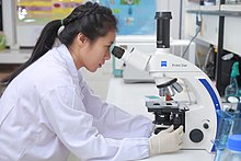
Optical microscope used at the Wiki Science Competition 2017 in Thailand |
|
| Uses | Small sample observation |
|---|---|
| Notable experiments | Discovery of cells |
| Related items | Optical microscope Electron microscope |
A microscope (from Ancient Greek μικρός (mikrós) ‘small’, and σκοπέω (skopéō) ‘to look (at); examine, inspect’) is a laboratory instrument used to examine objects that are too small to be seen by the naked eye. Microscopy is the science of investigating small objects and structures using a microscope. Microscopic means being invisible to the eye unless aided by a microscope.
There are many types of microscopes, and they may be grouped in different ways. One way is to describe the method an instrument uses to interact with a sample and produce images, either by sending a beam of light or electrons through a sample in its optical path, by detecting photon emissions from a sample, or by scanning across and a short distance from the surface of a sample using a probe. The most common microscope (and the first to be invented) is the optical microscope, which uses lenses to refract visible light that passed through a thinly sectioned sample to produce an observable image. Other major types of microscopes are the fluorescence microscope, electron microscope (both the transmission electron microscope and the scanning electron microscope) and various types of scanning probe microscopes.[1]
History
Although objects resembling lenses date back 4,000 years and there are Greek accounts of the optical properties of water-filled spheres (5th century BC) followed by many centuries of writings on optics, the earliest known use of simple microscopes (magnifying glasses) dates back to the widespread use of lenses in eyeglasses in the 13th century.[2][3][4] The earliest known examples of compound microscopes, which combine an objective lens near the specimen with an eyepiece to view a real image, appeared in Europe around 1620.[5] The inventor is unknown, even though many claims have been made over the years. Several revolve around the spectacle-making centers in the Netherlands, including claims it was invented in 1590 by Zacharias Janssen (claim made by his son) or Zacharias’ father, Hans Martens, or both,[6][7] claims it was invented by their neighbor and rival spectacle maker, Hans Lippershey (who applied for the first telescope patent in 1608),[8] and claims it was invented by expatriate Cornelis Drebbel, who was noted to have a version in London in 1619.[9][10] Galileo Galilei (also sometimes cited as compound microscope inventor) seems to have found after 1610 that he could close focus his telescope to view small objects and, after seeing a compound microscope built by Drebbel exhibited in Rome in 1624, built his own improved version.[11][12][13] Giovanni Faber coined the name microscope for the compound microscope Galileo submitted to the Accademia dei Lincei in 1625[14] (Galileo had called it the occhiolino ‘little eye’).
Rise of modern light microscopes

Carl Zeiss binocular compound microscope, 1914
The first detailed account of the microscopic anatomy of organic tissue based on the use of a microscope did not appear until 1644, in Giambattista Odierna’s L’occhio della mosca, or The Fly’s Eye.[15]
The microscope was still largely a novelty until the 1660s and 1670s when naturalists in Italy, the Netherlands and England began using them to study biology. Italian scientist Marcello Malpighi, called the father of histology by some historians of biology, began his analysis of biological structures with the lungs. The publication in 1665 of Robert Hooke’s Micrographia had a huge impact, largely because of its impressive illustrations. A significant contribution came from Antonie van Leeuwenhoek who achieved up to 300 times magnification using a simple single lens microscope. He sandwiched a very small glass ball lens between the holes in two metal plates riveted together, and with an adjustable-by-screws needle attached to mount the specimen.[16] Then, Van Leeuwenhoek re-discovered red blood cells (after Jan Swammerdam) and spermatozoa, and helped popularise the use of microscopes to view biological ultrastructure. On 9 October 1676, van Leeuwenhoek reported the discovery of micro-organisms.[15]
The performance of a light microscope depends on the quality and correct use of the condensor lens system to focus light on the specimen and the objective lens to capture the light from the specimen and form an image.[5] Early instruments were limited until this principle was fully appreciated and developed from the late 19th to very early 20th century, and until electric lamps were available as light sources. In 1893 August Köhler developed a key principle of sample illumination, Köhler illumination, which is central to achieving the theoretical limits of resolution for the light microscope. This method of sample illumination produces even lighting and overcomes the limited contrast and resolution imposed by early techniques of sample illumination. Further developments in sample illumination came from the discovery of phase contrast by Frits Zernike in 1953, and differential interference contrast illumination by Georges Nomarski in 1955; both of which allow imaging of unstained, transparent samples.
Electron microscopes

In the early 20th century a significant alternative to the light microscope was developed, an instrument that uses a beam of electrons rather than light to generate an image. The German physicist, Ernst Ruska, working with electrical engineer Max Knoll, developed the first prototype electron microscope in 1931, a transmission electron microscope (TEM). The transmission electron microscope works on similar principles to an optical microscope but uses electrons in the place of light and electromagnets in the place of glass lenses. Use of electrons, instead of light, allows for much higher resolution.
Development of the transmission electron microscope was quickly followed in 1935 by the development of the scanning electron microscope by Max Knoll.[17] Although TEMs were being used for research before WWII, and became popular afterwards, the SEM was not commercially available until 1965.
Transmission electron microscopes became popular following the Second World War. Ernst Ruska, working at Siemens, developed the first commercial transmission electron microscope and, in the 1950s, major scientific conferences on electron microscopy started being held. In 1965, the first commercial scanning electron microscope was developed by Professor Sir Charles Oatley and his postgraduate student Gary Stewart, and marketed by the Cambridge Instrument Company as the «Stereoscan».
One of the latest discoveries made about using an electron microscope is the ability to identify a virus.[18] Since this microscope produces a visible, clear image of small organelles, in an electron microscope there is no need for reagents to see the virus or harmful cells, resulting in a more efficient way to detect pathogens.
Scanning probe microscopes
From 1981 to 1983 Gerd Binnig and Heinrich Rohrer worked at IBM in Zurich, Switzerland to study the quantum tunnelling phenomenon. They created a practical instrument, a scanning probe microscope from quantum tunnelling theory, that read very small forces exchanged between a probe and the surface of a sample. The probe approaches the surface so closely that electrons can flow continuously between probe and sample, making a current from surface to probe. The microscope was not initially well received due to the complex nature of the underlying theoretical explanations. In 1984 Jerry Tersoff and D.R. Hamann, while at AT&T’s Bell Laboratories in Murray Hill, New Jersey began publishing articles that tied theory to the experimental results obtained by the instrument. This was closely followed in 1985 with functioning commercial instruments, and in 1986 with Gerd Binnig, Quate, and Gerber’s invention of the atomic force microscope, then Binnig’s and Rohrer’s Nobel Prize in Physics for the SPM.[19]
New types of scanning probe microscope have continued to be developed as the ability to machine ultra-fine probes and tips has advanced.
Fluorescence microscopes
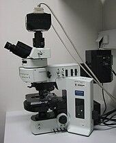
Fluorescence microscope with the filter cube turret above the objective lenses, coupled with a camera.
The most recent developments in light microscope largely centre on the rise of fluorescence microscopy in biology.[20] During the last decades of the 20th century, particularly in the post-genomic era, many techniques for fluorescent staining of cellular structures were developed.[20] The main groups of techniques involve targeted chemical staining of particular cell structures, for example, the chemical compound DAPI to label DNA, use of antibodies conjugated to fluorescent reporters, see
immunofluorescence, and fluorescent proteins, such as green fluorescent protein.[21] These techniques use these different fluorophores for analysis of cell structure at a molecular level in both live and fixed samples.
The rise of fluorescence microscopy drove the development of a major modern microscope design, the confocal microscope. The principle was patented in 1957 by Marvin Minsky, although laser technology limited practical application of the technique. It was not until 1978 when Thomas and Christoph Cremer developed the first practical confocal laser scanning microscope and the technique rapidly gained popularity through the 1980s.
Super resolution microscopes
Much current research (in the early 21st century) on optical microscope techniques is focused on development of superresolution analysis of fluorescently labelled samples. Structured illumination can improve resolution by around two to four times and techniques like stimulated emission depletion (STED) microscopy are approaching the resolution of electron microscopes.[22] This occurs because the diffraction limit is occurred from light or excitation, which makes the resolution must be doubled to become super saturated. Stefan Hell was awarded the 2014 Nobel Prize in Chemistry for the development of the STED technique, along with Eric Betzig and William Moerner who adapted fluorescence microscopy for single-molecule visualization.[23]
X-ray microscopes
X-ray microscopes are instruments that use electromagnetic radiation usually in the soft X-ray band to image objects. Technological advances in X-ray lens optics in the early 1970s made the instrument a viable imaging choice.[24] They are often used in tomography (see micro-computed tomography) to produce three dimensional images of objects, including biological materials that have not been chemically fixed. Currently research is being done to improve optics for hard X-rays which have greater penetrating power.[24]
Types
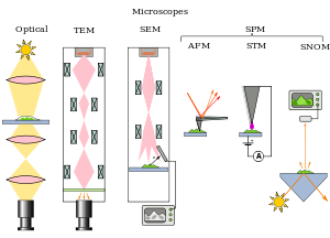
Types of microscopes illustrated by the principles of their beam paths

Evolution of spatial resolution achieved with optical, transmission (TEM) and aberration-corrected electron microscopes (ACTEM).[25]
Microscopes can be separated into several different classes. One grouping is based on what interacts with the sample to generate the image, i.e., light or photons (optical microscopes), electrons (electron microscopes) or a probe (scanning probe microscopes). Alternatively, microscopes can be classified based on whether they analyze the sample via a scanning point (confocal optical microscopes, scanning electron microscopes and scanning probe microscopes) or analyze the sample all at once (wide field optical microscopes and transmission electron microscopes).
Wide field optical microscopes and transmission electron microscopes both use the theory of lenses (optics for light microscopes and electromagnet lenses for electron microscopes) in order to magnify the image generated by the passage of a wave transmitted through the sample, or reflected by the sample. The waves used are electromagnetic (in optical microscopes) or electron beams (in electron microscopes). Resolution in these microscopes is limited by the wavelength of the radiation used to image the sample, where shorter wavelengths allow for a higher resolution.[20]
Scanning optical and electron microscopes, like the confocal microscope and scanning electron microscope, use lenses to focus a spot of light or electrons onto the sample then analyze the signals generated by the beam interacting with the sample. The point is then scanned over the sample to analyze a rectangular region. Magnification of the image is achieved by displaying the data from scanning a physically small sample area on a relatively large screen. These microscopes have the same resolution limit as wide field optical, probe, and electron microscopes.
Scanning probe microscopes also analyze a single point in the sample and then scan the probe over a rectangular sample region to build up an image. As these microscopes do not use electromagnetic or electron radiation for imaging they are not subject to the same resolution limit as the optical and electron microscopes described above.
Optical microscope
The most common type of microscope (and the first invented) is the optical microscope. This is an optical instrument containing one or more lenses producing an enlarged image of a sample placed in the focal plane. Optical microscopes have refractive glass (occasionally plastic or quartz), to focus light on the eye or on to another light detector. Mirror-based optical microscopes operate in the same manner. Typical magnification of a light microscope, assuming visible range light, is up to 1,250× with a theoretical resolution limit of around 0.250 micrometres or 250 nanometres.[20] This limits practical magnification to ~1,500×. Specialized techniques (e.g., scanning confocal microscopy, Vertico SMI) may exceed this magnification but the resolution is diffraction limited. The use of shorter wavelengths of light, such as ultraviolet, is one way to improve the spatial resolution of the optical microscope, as are devices such as the near-field scanning optical microscope.
Sarfus is a recent optical technique that increases the sensitivity of a standard optical microscope to a point where it is possible to directly visualize nanometric films (down to 0.3 nanometre) and isolated nano-objects (down to 2 nm-diameter). The technique is based on the use of non-reflecting substrates for cross-polarized reflected light microscopy.
Ultraviolet light enables the resolution of microscopic features as well as the imaging of samples that are transparent to the eye. Near infrared light can be used to visualize circuitry embedded in bonded silicon devices, since silicon is transparent in this region of wavelengths.
In fluorescence microscopy many wavelengths of light ranging from the ultraviolet to the visible can be used to cause samples to fluoresce, which allows viewing by eye or with specifically sensitive cameras.

Unstained cells viewed by typical brightfield (left) compared to phase-contrast microscopy (right).
Phase-contrast microscopy is an optical microscopic illumination technique in which small phase shifts in the light passing through a transparent specimen are converted into amplitude or contrast changes in the image.[20] The use of phase contrast does not require staining to view the slide. This microscope technique made it possible to study the cell cycle in live cells.
The traditional optical microscope has more recently evolved into the digital microscope. In addition to, or instead of, directly viewing the object through the eyepieces, a type of sensor similar to those used in a digital camera is used to obtain an image, which is then displayed on a computer monitor. These sensors may use CMOS or charge-coupled device (CCD) technology, depending on the application.
Digital microscopy with very low light levels to avoid damage to vulnerable biological samples is available using sensitive photon-counting digital cameras. It has been demonstrated that a light source providing pairs of entangled photons may minimize the risk of damage to the most light-sensitive samples. In this application of ghost imaging to photon-sparse microscopy, the sample is illuminated with infrared photons, each of which is spatially correlated with an entangled partner in the visible band for efficient imaging by a photon-counting camera.[26]
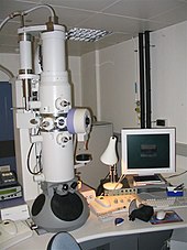
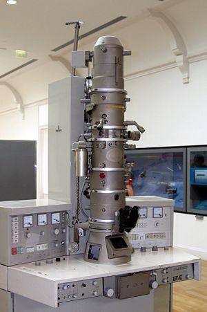
Modern transmission electron microscope
Electron microscope
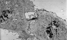
Transmission electron micrograph of a dividing cell undergoing cytokinesis
The two major types of electron microscopes are transmission electron microscopes (TEMs) and scanning electron microscopes (SEMs).[20][21] They both have series of electromagnetic and electrostatic lenses to focus a high energy beam of electrons on a sample. In a TEM the electrons pass through the sample, analogous to basic optical microscopy.[20] This requires careful sample preparation, since electrons are scattered strongly by most materials.[21] The samples must also be very thin (below 100 nm) in order for the electrons to pass through it.[20][21] Cross-sections of cells stained with osmium and heavy metals reveal clear organelle membranes and proteins such as ribosomes.[21] With a 0.1 nm level of resolution, detailed views of viruses (20 – 300 nm) and a strand of DNA (2 nm in width) can be obtained.[21] In contrast, the SEM has raster coils to scan the surface of bulk objects with a fine electron beam. Therefore, the specimen do not necessarily need to be sectioned, but coating with a nanometric metal or carbon layer may be needed for nonconductive samples.[20] SEM allows fast surface imaging of samples, possibly in thin water vapor to prevent drying.[20][21]
Scanning probe
The different types of scanning probe microscopes arise from the many different types of interactions that occur when a small probe is scanned over and interacts with a specimen. These interactions or modes can be recorded or mapped as function of location on the surface to form a characterization map. The three most common types of scanning probe microscopes are atomic force microscopes (AFM), near-field scanning optical microscopes (MSOM or SNOM, scanning near-field optical microscopy), and scanning tunneling microscopes (STM).[27] An atomic force microscope has a fine probe, usually of silicon or silicon nitride, attached to a cantilever; the probe is scanned over the surface of the sample, and the forces that cause an interaction between the probe and the surface of the sample are measured and mapped. A near-field scanning optical microscope is similar to an AFM but its probe consists of a light source in an optical fiber covered with a tip that has usually an aperture for the light to pass through. The microscope can capture either transmitted or reflected light to measure very localized optical properties of the surface, commonly of a biological specimen. Scanning tunneling microscopes have a metal tip with a single apical atom; the tip is attached to a tube through which a current flows.[28] The tip is scanned over the surface of a conductive sample until a tunneling current flows; the current is kept constant by computer movement of the tip and an image is formed by the recorded movements of the tip.[27]
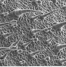
Leaf surface viewed by a scanning electron microscope.
Other types
Scanning acoustic microscopes use sound waves to measure variations in acoustic impedance. Similar to Sonar in principle, they are used for such jobs as detecting defects in the subsurfaces of materials including those found in integrated circuits. On February 4, 2013, Australian engineers built a «quantum microscope» which provides unparalleled precision.[29]
Mobile apps
Mobile app microscopes can optionally be used as optical microscope when the flashlight is activated. However, mobile app microscopes are harder to use due to visual noise, are often limited to 40x, and the resolution limits of the camera lens itself.
See also
- Fluorescence interference contrast microscopy
- Laser capture microdissection
- Microscope image processing
- Microscope slide
- Multifocal plane microscopy
- Royal Microscopical Society
References

First atomic force microscope
- ^ Characterization and Analysis of Polymers. Hoboken, NJ: Wiley-Interscience. 2008. ISBN 978-0-470-23300-9.
- ^ Bardell, David (May 2004). «The Invention of the Microscope». BIOS. 75 (2): 78–84. doi:10.1893/0005-3155(2004)75<78:tiotm>2.0.co;2. JSTOR 4608700. S2CID 96668398.
- ^ The history of the telescope by Henry C. King, Harold Spencer Jones Publisher Courier Dover Publications, 2003, pp. 25–27 ISBN 0-486-43265-3, 978-0-486-43265-6
- ^ Atti Della Fondazione Giorgio Ronchi E Contributi Dell’Istituto Nazionale Di Ottica, Volume 30, La Fondazione-1975, p. 554
- ^ a b Murphy, Douglas B.; Davidson, Michael W. (2011). Fundamentals of light microscopy and electronic imaging (2nd ed.). Oxford: Wiley-Blackwell. ISBN 978-0-471-69214-0.
- ^ Sir Norman Lockyer (1876). Nature Volume 14.
- ^ Albert Van Helden; Sven Dupré; Rob van Gent (2010). The Origins of the Telescope. Amsterdam University Press. pp. 32–36, 43. ISBN 978-90-6984-615-6.
- ^ «Who Invented the Microscope?». Live Science. 14 September 2013. Retrieved 31 March 2017.
- ^ Eric Jorink (2010-10-25). Reading the Book of Nature in the Dutch Golden Age, 1575-1715. ISBN 978-90-04-18671-2.
- ^ William Rosenthal, Spectacles and Other Vision Aids: A History and Guide to Collecting, Norman Publishing, 1996, pp. 391–92
- ^ Raymond J. Seeger, Men of Physics: Galileo Galilei, His Life and His Works, Elsevier – 2016, p. 24
- ^ J. William Rosenthal, Spectacles and Other Vision Aids: A History and Guide to Collecting, Norman Publishing, 1996, page 391
- ^ uoregon.edu, Galileo Galilei (Excerpt from the Encyclopedia Britannica)
- ^ Gould, Stephen Jay (2000). «Chapter 2: The Sharp-Eyed Lynx, Outfoxed by Nature». The Lying Stones of Marrakech: Penultimate Reflections in Natural History. New York: Harmony. ISBN 978-0-224-05044-9.
- ^ a b Wootton, David (2006). Bad medicine: doctors doing harm since Hippocrates. Oxford [Oxfordshire]: Oxford University Press. p. 110. ISBN 978-0-19-280355-9.[page needed]
- ^ Liz Logan (27 April 2016). «Early Microscopes Revealed a New World of Tiny Living Things». Smithsonian.com. Retrieved 3 June 2016.
- ^ Knoll, Max (1935). «Aufladepotentiel und Sekundäremission elektronenbestrahlter Körper». Zeitschrift für Technische Physik. 16: 467–475.
- ^ Goldsmith, Cynthia S.; Miller, Sara E. (2009-10-01). «Modern Uses of Electron Microscopy for Detection of Viruses». Clinical Microbiology Reviews. 22 (4): 552–563. doi:10.1128/cmr.00027-09. ISSN 0893-8512. PMC 2772359. PMID 19822888.
- ^ Morita, Seizo (2007). Roadmap of Scanning Probe Microscopy. Berlin, Heidelberg: Springer-Verlag Berlin Heidelberg. ISBN 978-3-540-34315-8.
- ^ a b c d e f g h i j Lodish, Harvey; Berk, Arnold; Zipursky, S. Lawrence; Matsudaira, Paul; Baltimore, David; Darnell, James (2000). «Microscopy and Cell Architecture». Molecular Cell Biology. 4th Edition.
- ^ a b c d e f g Alberts, Bruce; Johnson, Alexander; Lewis, Julian; Raff, Martin; Roberts, Keith; Walter, Peter (2002). «Looking at the Structure of Cells in the Microscope». Molecular Biology of the Cell. 4th Edition.
- ^ «The Nobel Prize in Chemistry 2014 – Scientific Background» (PDF). www.nobelprize.org. Retrieved 2018-03-20.
- ^ «The Nobel Prize in Chemistry 2014». www.nobelprize.org. Retrieved 2018-03-20.
- ^ a b Erko, A. (2008). Modern developments in X-ray and neutron optics. Berlin: Springer. ISBN 978-3-540-74561-7.
- ^ Pennycook, S.J.; Varela, M.; Hetherington, C.J.D.; Kirkland, A.I. (2011). «Materials Advances through Aberration-Corrected Electron Microscopy» (PDF). MRS Bulletin. 31: 36–43. doi:10.1557/mrs2006.4.
- ^ Aspden, Reuben S.; Gemmell, Nathan R.; Morris, Peter A.; Tasca, Daniel S.; Mertens, Lena; Tanner, Michael G.; Kirkwood, Robert A.; Ruggeri, Alessandro; Tosi, Alberto; Boyd, Robert W.; Buller, Gerald S.; Hadfield, Robert H.; Padgett, Miles J. (2015). «Photon-sparse microscopy: visible light imaging using infrared illumination» (PDF). Optica. 2 (12): 1049. Bibcode:2015Optic…2.1049A. doi:10.1364/OPTICA.2.001049. ISSN 2334-2536.
- ^ a b Bhushan, Bharat, ed. (2010). Springer handbook of nanotechnology (3rd rev. & extended ed.). Berlin: Springer. p. 620. ISBN 978-3-642-02525-9.
- ^ Sakurai, T.; Watanabe, Y., eds. (2000). Advances in scanning probe microscopy. Berlin: Springer. ISBN 978-3-642-56949-4.
- ^ «Quantum Microscope for Living Biology». Science Daily. 4 February 2013. Retrieved 5 February 2013.
External links
- Milestones in Light Microscopy, Nature Publishing
- FAQ on Optical Microscopes
- Nikon MicroscopyU, tutorials from Nikon
- Molecular Expressions : Exploring the World of Optics and Microscopy, Florida State University.
What is microscope word?
1 : an optical instrument consisting of a lens or combination of lenses for making enlarged images of minute objects especially : compound microscope. 2 : a non-optical instrument (such as one using radiations other than light or using vibrations) for making enlarged images of minute objects an acoustic microscope.
What is the root word of scientist?
The noun scientist comes from Latin ‘scientia’, probably via the stem of the adjective ‘scientific’ plus the suffix -ist. It was first recorded in English in the first part of the 19th century and has replaced the earlier term ‘sciencist’.
What is a scientist in one word?
1 : a person learned in science and especially natural science : a scientific investigator. 2 capitalized : christian scientist.
What is Isscientist?
A scientist is a person who conducts scientific research to advance knowledge in an area of interest. In classical antiquity, there was no real ancient analog of a modern scientist. Instead, philosophers engaged in the philosophical study of nature called natural philosophy, a precursor of natural science.
Do you call a scientist Dr?
Scientists who are called “Doctor” are not medical doctors, like the ones who take care of you when you’re sick. Their title refers to the level of specialization and education that they have achieved in their field of study.
Is a psychologist a professional?
Psychologists are registered health professionals who are highly trained to support individuals, families, careers and other health professionals in palliative care settings.
What is the title for a psychologist?
Under CA law, “psychologist” is a protected job title. A person can use this title only if he or she has obtained a doctoral degree in psychology: a PhD, PsyD, or EdD. (The differences between these degrees is explained in the section of this website called Doctoral Degrees for Psychologists).
Do you need a PHD to be called a psychologist?
How long does it take to become a licensed psychologist in California? To become a licensed psychologist in California, you need to have a doctorate degree, which can take up to 10 years to obtain. You will also have to acquire 3,000 hours of supervised experience, 1,500 which can be pre-doctoral.
Can you call yourself a psychologist with a master’s degree?
While many institutions offer bachelor’s degrees in psychology, in order to actually call yourself a “psychologist,” you’ll need at least a master’s degree, and in most cases you’ll need a doctorate. Opportunities will be limited for those who hold bachelor’s degrees or less in psychology.
![]()
![]()
- Entertainment & Pop Culture
- Geography & Travel
- Health & Medicine
- Lifestyles & Social Issues
- Literature
- Philosophy & Religion
- Politics, Law & Government
- Science
- Sports & Recreation
- Technology
- Visual Arts
- World History
- On This Day in History
- Quizzes
- Podcasts
- Dictionary
- Biographies
- Summaries
- Top Questions
- Infographics
- Demystified
- Lists
- #WTFact
- Companions
- Image Galleries
- Spotlight
- The Forum
- One Good Fact
- Entertainment & Pop Culture
- Geography & Travel
- Health & Medicine
- Lifestyles & Social Issues
- Literature
- Philosophy & Religion
- Politics, Law & Government
- Science
- Sports & Recreation
- Technology
- Visual Arts
- World History
- Britannica Explains
In these videos, Britannica explains a variety of topics and answers frequently asked questions. - Britannica Classics
Check out these retro videos from Encyclopedia Britannica’s archives. - Demystified Videos
In Demystified, Britannica has all the answers to your burning questions. - #WTFact Videos
In #WTFact Britannica shares some of the most bizarre facts we can find. - This Time in History
In these videos, find out what happened this month (or any month!) in history.
- Student Portal
Britannica is the ultimate student resource for key school subjects like history, government, literature, and more. - COVID-19 Portal
While this global health crisis continues to evolve, it can be useful to look to past pandemics to better understand how to respond today. - 100 Women
Britannica celebrates the centennial of the Nineteenth Amendment, highlighting suffragists and history-making politicians. - Saving Earth
Britannica Presents Earth’s To-Do List for the 21st Century. Learn about the major environmental problems facing our planet and what can be done about them! - SpaceNext50
Britannica presents SpaceNext50, From the race to the Moon to space stewardship, we explore a wide range of subjects that feed our curiosity about space!
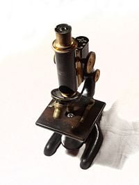
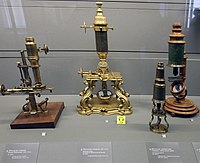
A 1915 Bausch and Lomb microscope
A microscope (Greek: μικρόν (micron) = small + σκοπεῖν (skopein) = to look at) is an instrument for viewing objects that are too small to be seen by the naked or unaided eye. Microscopes give us a larger (magnified) image of a tiny object. The microscopes we use in school and at home trace their history back almost four hundred years.
The most common type of microscope—and the first to be invented—is the optical microscope. This is an optical instrument containing one or more lenses that produce an enlarged image of an object placed in the focal plane of the lens(es). However, human creativity, employed in exploring nature, manifested in numerous improvements and new microscope designs, including the electron microscope, scanning probe microscope, field ion microscope, acoustic microscope, and so forth.
The science of investigating small objects using such an instrument is called microscopy, and the term microscopic means minute or very small, not easily visible with the unaided eye; in other words, requiring a microscope to examine. Microbiology is the scientific study of microorganisms, which are forms of life that are microscopic, such as bacteria, fungi, archaea, and protists.
Microscopes extending the range of human visual sensing reveal a world much grander than the world visible to the unaided eye and remind us of how much we cannot know by our own senses. As new microscopes have revealed new layers of the microscopic world successively further and further removed from the human scale, they have raised new challenges of understanding because objects at different scales have their own distinctive behavior properties, sometimes quite different than those at the human scale. When a scanning tunneling microscope, for example, maps the surface of a crystal and shows us a neat array of fuzzy atoms we can get a better visualization of how the atoms may be arranged, but we also need to remember that those fuzzy balls comprise primarily empty space and that we, ourselves, are no more solid at the atomic level than those fuzzy balls in the picture.
History
The first useful microscope is considered to have been developed in the Netherlands in the late sixteenth and early seventeenth century. There is almost as much confusion about the inventor as about the dates. Three different eyeglass makers have been given credit for the invention: Hans Lippershey (who also developed the first real telescope); Hans Janssen; and his son, Zacharias Janssen. Hans Janssen and Zacharias are often said to have invented the first compound microscope in 1590, but this was a declaration by Zacharias Janssen himself halfway through the seventeenth century. The date is certainly not likely, as it has been shown that Zacharias Janssen actually was born around 1590.
By 1609 Galileo Galilei is also credited with inventing a compound microscope with a convex and a concave lens, which he presented to Polish king Sigismund III in 1612.
In 1625 Giovanni Faber of Bamberg (1574–1629) coined the word microscope by analogy with telescope.
Lens quality in early microscopes was often poor so the images were not very clear. But even these rather crude microscopes were a great help in learning more about animals and plants. For example, in 1665, Robert Hooke (1635–1703) published Micrographia, a collection of biological micrographs developed using the compound microscope. Hooked coined the word cell for the structures he discovered in cork bark.
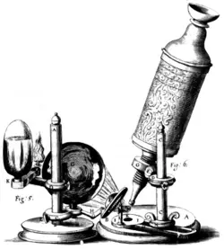
Thonius Philips van Leeuwenhoek, better known as Anton van Leeuwenhoek (1632–1723), was a Dutch tradesman who is well known for his contribution to the improvement of the microscope, as well as his contributions toward the establishment of microbiology. Known as «the father of microbiology,» Leeuwenhoek, using his handcrafted microscopes, was the first to observe and describe single celled organisms that he first referred to as animalcules, and which we now refer to as microorganisms.
Leeuwenhoek’s interest in microscopes began in 1648 in Amsterdam, when he saw a simple microscope, a magnifying glass mounted on a small stand used by textile merchants capable of magnifying to a power of three. It is believed that soon after 1665, he read Robert Hooke’s book Micrographia, and this roused an interest in using microscopes for the purpose of investigating the natural world.
Leeuwenhoek’s interest in microscopy grew steadily until he was spending most of his nights and free time grinding his own lenses, improving the quality of his microscopes, and studying everything he could beneath them. Although he is sometimes erroneously referred to as «the inventor of the microscope,» compound microscopes (with two lenses mounted together) had existed for about three-quarters of a century. However, they were very crude because the technology used made it difficult to build them properly. Leeuwenhoek’s genius was in developing his skill to grind single lenses very precisely. It is likely that his microscopes were powerful magnifying glasses, not compound microscopes.
During his lifetime Leeuwenhoek ground over five hundred optical lenses. He also created over four hundred different types of microscopes, nine of which still exist today. His microscopes were made of silver or copper metal frames holding hand-ground lenses. Those that survived the years are able to magnify up to 270 times. It is suspected, though, that Leeuwenhoek possessed some microscopes that could magnify up to five hundred times.
In the 1860s, Ernst Abbe discovered the Abbe sine condition, a mathematical condition that must be fulfilled by a lens or other optical system in order for it to produce sharp images of off-axis as well as on-axis objects. This offered a breakthrough in microscope design, which until then was largely based on trial and error. The company of Carl Zeiss exploited this discovery and became the dominant microscope manufacturer of its era.
In 1931 Ernst Ruska started to build the first electron microscope, a transmission electron microscope (TEM). In 1936 Erwin Müller invented the field emission microscope, and in 1951 he invented the field ion microscope and was the first to see atoms. In 1967 he added time-of-flight spectroscopy to the field ion microscope, making the first atom probe and allowing the chemical identification of each individual atom.
In 1953 Frits Zernike, professor of theoretical physics, received the Nobel Prize in Physics for his invention of the phase contrast microscope.
In 1981 Gerd Binnig and Heinrich Rohrer developed the scanning tunneling microscope, and in 1986, Binnig, Quate, and Gerber invented the atomic force microscope. In 1988 Alfred Cerezo, Terence Godfrey, and George Smith applied a position-sensitive detector to the atom probe, making it able to resolve atoms in 3-dimensions. Also in 1988 Kingo Itaya invented the electrochemical scanning tunneling microscope, and in 1991, the Kelvin probe force microscope was invented.
Microscope Types
Microscopes can largely be separated into two classes, optical theory microscopes and scanning probe microscopes.
Optical theory microscopes are microscopes that function through the optical theory of lenses in order to magnify the image generated by the passage of a wave through the sample. The waves used are either electromagnetic in optical microscopes or electron beams in electron microscopes. The types commonly used today are the compound light microscope, stereo microscope, and the electron microscope.
Scanning probe microscopes form images of surfaces using a physical probe that scans the specimen. An image of the surface is obtained by mechanically moving the probe in a raster scan of the specimen, line by line, and recording the probe-surface interaction as a function of position.
Optical microscopes
A stereo microscope is often used for lower-power magnification on large subjects
The optical microscope is a type of microscope that uses visible light and a system of lenses to magnify images of small samples. Optical microscopes, through their use of visible wavelengths of light, are the simplest and hence most widely used type of microscope.
There are two basic configurations of optical microscope in use, the simple microscope that uses only one lens for magnification, and the compound microscope that uses a set of many lenses in order to maximize magnification. Recent research [1] has shown that even simple microscopes, those with a single small lens, gave amazingly clear images to the earliest microscopists.
The compound light microscope, in its simplest form—as used by Hooke, for example—would have a single glass lens of short focal length for the objective, and another single glass lens for the eyepiece or ocular. Modern microscopes of this kind are usually more complex, with multiple lens components in both objective and eyepiece assemblies. Today, compound microscopes serve uses in many fields of science, particularly biology and geology.
Optical microscopes use refractive lenses, typically of glass and occasionally of plastic, to focus light into the eye or another light detector. Typical magnification of a light microscope is up to 1,500 times with a theoretical resolution of around 0.2 micrometers. Specialized techniques (e.g., scanning confocal microscopy) may exceed this magnification, but the resolution is an insurmountable diffraction limit.
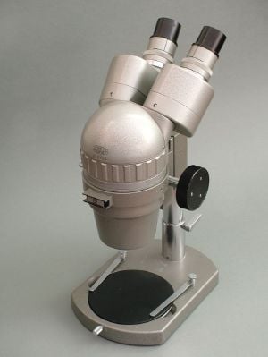
The stereo or dissecting microscope uses two separate optical paths with two objectives and two eyepieces to provide slightly different viewing angles to the left and right eyes. In this way it produces a three-dimensional (3-D) visualization of the sample being examined. The stereo microscope is often used to study the surfaces of solid specimens or to carry out close work such as sorting, dissection, microsurgery, watch-making, small circuit board manufacture or inspection, and the like.
Electromagnetic wavelengths not visible to human eye
Other microscopes that use electromagnetic wavelengths not visible to the human eye are often called optical microscopes. The most common of these, due to its high resolution yet no requirement for a vacuum-like electron microscope, is the x-ray microscope.
Electron microscopes
Electron microscopes, which use beams of electrons instead of light, are designed for very high magnification usage. Electrons, which have a much smaller wavelength than visible light, allow a much higher resolution. The main limitation of the electron beam is that it must pass through a vacuum as air molecules would otherwise scatter the beam.
Instead of relying on refraction, lenses for electron microscopes are specially designed electromagnets that generates magnetic fields that are approximately parallel to the direction that electrons travel. The electrons are typically detected by a phosphor screen, photographic film or a charge-coupled device (CCD).
Two major variants of electron microscopes exist:
- Scanning electron microscope: looks at the surface of bulk objects by scanning the surface with a fine electron beam and measuring reflection. May also be used for spectroscopy.
- Transmission electron microscope: passes electrons completely through the sample, analogous to basic optical microscopy. This requires careful sample preparation, since electrons are scattered so strongly by most materials. It can also obtain detailed information on the sample’s crystallography through selected area diffraction.
Scanning probe microscope
In scanning probe microscopy (SPM), a physical probe is used either in close contact to the sample or nearly touching it. By rastering the probe across the sample, and by measuring the interactions between the sharp tip of the probe and the sample, a micrograph is generated. The exact nature of the interactions between the probe and the sample determines exactly what kind of SPM is being used. Because this kind of microscopy relies on the interactions between the tip and the sample, it generally only measures information about the surface of the sample.
Some kinds of SPMs are:
- Atomic force microscope
- Scanning tunneling microscope
- Electric force microscope
- Magnetic force microscope (MFM)
- Near-field scanning optical microscope
Point-projection microscopes
The field emission microscope, field ion microscope, and the Atom Probe are examples of point-projection microscopes, where ions are excited from a needle-shaped specimen and hit a detector. The Atom-Probe Tomograph (APT) is the most modern incarnation and allows a three-dimensional atom-by-atom (with chemical elements identified) reconstruction with sub-nanometer resolution.
Acoustic microscopes
Acoustic microscopes use sound waves to measure variations in acoustic impedance. Similar to SONAR in principle, they are used for such jobs as detecting defects in the subsurfaces of materials including those found in integrated circuits.
References
ISBN links support NWE through referral fees
- Dobell, C. (ed.) 1960. Antony van Leeuwenhoek and his «Little Animals. New York: Dover Publications.
- Ford, B. J. 1991. The Leeuwenhoek Legacy. London: Biopress, Bristol, and Farrand Press. ISBN 1850830169.
- Van Berkel, K. “Vermeer, Van Leeuwenhoek en De Astronoom.” Vrij Nederland (February 24, 1996): 62-67.
Credits
New World Encyclopedia writers and editors rewrote and completed the Wikipedia article
in accordance with New World Encyclopedia standards. This article abides by terms of the Creative Commons CC-by-sa 3.0 License (CC-by-sa), which may be used and disseminated with proper attribution. Credit is due under the terms of this license that can reference both the New World Encyclopedia contributors and the selfless volunteer contributors of the Wikimedia Foundation. To cite this article click here for a list of acceptable citing formats.The history of earlier contributions by wikipedians is accessible to researchers here:
- Microscope history
- Timeline_of_microscope_technology history
- Optical_microscope history
The history of this article since it was imported to New World Encyclopedia:
- History of «Microscope»
Note: Some restrictions may apply to use of individual images which are separately licensed.