
21. Используйте производные от слов справа для того, чтобы дописать текст.
Слово “микроскоп” представляет собой (1) сочетание двух греческих слов, micros, или “маленький”, и skopos, или “наблюдатель”. Итак, микроскоп — это наблюдатель малого. Он помогает увидеть крошечные вещи, которые (2) невидимы невооруженным глазом. Он был изобретен между 1590 и 1610. Никто не уверен, кто (3) на самом деле это сделал. (4) Обычно это считается заслугой Галилея. Голландский (5) ученый по имени Левенгук иногда упоминается как “отец микроскопа”, но это из-за многих (6) открытий, которые он сделал с этим инструментом. Он был первым, кто увидел такие (7) микроскопические формы жизни, как бактерии. Он также был первым, кто увидел всю (8) циркуляцию крови. Сегодня (9) значение микроскопа для человека почти во всех важных областях науки и промышленности очевидно.
ГДЗ #
1) combination (сочетание);
2) invisible (невидимы);
3) actually (на самом деле);
4) usually (обычно);
5) scientist (ученый);
6) discoveries (открытий);
7) microscopic (микроскопические);
 circulation (циркуляцию);
9) importance (значение).
21. Use the derivatives of the words on the right to complete the text.
The word microscope’ is a (1) combination of two Greek words, micros, or “small”, and skopos, or “watcher”. So a microscope is a watcher of the small. It helps to see tiny things which are (2) invisible to the naked eye. It was invented sometime between 1590 and 1610. No one is quite sure who (3) actually did it. The credit is (4) usually given to Galileo. A Dutch (5) scientist called Leeuwenhoek is sometimes referred to as “the father of microscope” but that’s because of many (6) discoveries he made with this instrument. He was the first to see such (7) microscopic forms of life as bacteria. He was also the first to see the whole (8) circulation of the blood. Today the circulate (9) importance of microscope to man in almost every important form of science and industry is evident.
На этой странице вы сможете найти и списать готовое домешнее задание (ГДЗ) для школьников по предмету Английский язык, которые посещают 11 класс из книги или рабочей тетради под названием/издательством «Решебник ГДЗ Rainbow English», которая была написана автором/авторами: Афанасьева. ГДЗ представлено для списывания совершенно бесплатно и в открытом доступе.
![]()
68 месяцев назад
how did he do it? 3)when was the first microscope built? 4)what were main characteristics of the first microscope made by Zacharias Jansenn? 5)who was the first man to see bacteria with help of microscope? what were the main characteristics of his microscope? 6)who was the first inventor of the telescope? what do historians say? 7)what were the main characteristics of the first telescope? 8)what made it possible to do with powerful telescopes? 9)what did the great Italian scientist Galileo Galilei discover with the help of telescopes?
Ответы
Будь первым, кто ответит на вопрос
Контрольная работа по английскому языку в 11 классе за 3 четверть. (вариант 1)
1. Read the text and complete it with the phrases (a-h). There is one phrase you don’t have to use.
a) surgeon operates the instruments
b) by an electronic device
c) the building of surgical robots
d) because they can detect the change in acidity
e) becomes more difficult
f) on a special screen
g) the operation can be done faster
h) safe are these robots
Now there are robots that help surgeon perform operations. What can they do and is there anything to fear?
As surgical techniques become more advanced, keeping close control over everything that goes on during the operation (1)_________. The need for greater control and sensitivity has led to (2)_________. Keyhole surgery has s lot of benefit from their help. Specially adapted instruments on the ends of thin tubes are put through a small hole in the patient’s skin. The (3)_________ from outside the body using an instrument called a laparoscope, which has a camera on it. Thus doctors can watch the whole operation (4)_________.
The Laparobot or the robot laparascope gives the surgeon greater control over the image on the screen, so (5)_________. The Laparobot is controlled (6)_________ worn by the surgeon. The position of the camera inside the patient follows the surgeon’s hands movements. But how (7)_________ ? Unlike industrial robots, these new surgical robots are designed to be extremely sensitive to the environment and respond quickly to any change. For instance, they stop if they bump into an unexpected blood vessel.
2. Translate into English.
1. изумительный 2. захватывать 3. понимать 4. убедительный 5. тонуть (о людях) 6. выставлять 7. верный 8. роскошный 9. обладать 10. выпускать 11.искать 12. подходящий 13. подозревать 14. стоящий 15. придираться
Контрольная работа по английскому языку в 11 классе за 3 четверть. (вариант 2)
1. Read the text and complete it with the phrases (a-h). There is one phrase you don’t have to use.
a) safe are these robots
b) the operation can be done faster
c) becomes more difficult
d) on a special screen
e) the building of surgical robots
f) by an electronic device
g) because they can detect the change in acidity
h) surgeon operates the instruments
Now there are robots that help surgeon perform operations. What can they do and is there anything to fear?
As surgical techniques become more advanced, keeping close control over everything that goes on during the operation (1)_________. The need for greater control and sensitivity has led to (2)_________. Keyhole surgery has s lot of benefit from their help. Specially adapted instruments on the ends of thin tubes are put through a small hole in the patient’s skin. The (3)_________ from outside the body using an instrument called a laparoscope, which has a camera on it. Thus doctors can watch the whole operation (4)_________.
The Laparobot or the robot laparascope gives the surgeon greater control over the image on the screen, so (5)_________. The Laparobot is controlled (6)_________ worn by the surgeon. The position of the camera inside the patient follows the surgeon’s hands movements. But how (7)_________ ? Unlike industrial robots, these new surgical robots are designed to be extremely sensitive to the environment and respond quickly to any change. For instance, they stop if they bump into an unexpected blood vessel.
2. Translate into English.
1. изумление 2. захватывающий 3. понятный 4. убеждать 5. тонуть (о неодушевленных предметах) 6. выставка 7. вера 8. роскошь 9. обладание 10. восстанавливать 11.напоминать 12. подходить 13. подозрение 14.быть уволенным 15. выбирать
3. Match the words in two columns and complete the sentences with the phrases.
1. hardly a) a gardener
2. worth b) the poor animal
3. employ c) of restoring
4. suspected d) of being a spy
5. released e) comprehensible
1. She was ___________ but the police didn’t have any evidence. 2. Sir Reynolds was reading a long ___________ report written in official language. 3. The old church is certainly ___________ but the community doesn’t have any money to pay the architect and the builders. 4. The new owner of the castle intended to ___________ and the cook. 5. Walking in the forest Sam saw a small white hare under a thick branch and ___________ from the trap.
4. Use the adverbs in brackets in the appropriate forms to complete the sentences.
1. Two very large vans drove quite slowly down the street. The first one was moving a bit (quickly) than the second one. 2. The last orator spoke (enthusiastically) in comparison to the previous speakers. 3. The diggers moved much (far) but didn’t reach the bottom. 4. I see Uncle Gerald (little) of all my relatives because he seldom comes to Wales. 5.Such species appear (frequently) here than the ones we were speaking about the other day. 6. She began to smile (brightly) than in the days of his youth.
5. Use the derivatives of the words given in brackets to complete the text.
The word “microscope” is a 1.(combine) of two Greek words, micros, or “small” and scopos, or “watcher”. It helps to see tiny things, which are 2.(visible) to the naked eye. No one is quite sure who 3.(actual) invented it. The credit is 4.(usual) given to Galileo. A Dutch 5(science) called Leeuwenhoek is sometimes referred to as “the father of microscope’ but that’s because of many 6(discover) he made with this instrument. He was also the first to see the whole 7(circulate) of the blood. Today the 8(important) of microscope to man in almost every form of science and industry is evident.
3. Match the words in two columns and complete the sentences with the phrases.
1. masterpiece a) scolar
2. famous b) beauty
3. captivating c) a car
4. perform d) miracles
5. hired e) is on exhibition
1. In his book “English as a global language” David Cristal, a ___________ and a linguist, showed the role of English in the modern world. 2. Sunflowers Vincent Van Gogh’s ___________in Sweden. 3. Every year Venice’s ___________ attracts thousands of tourists. 4. They say Doctor Lewis can really ___________. Practically all his patients leave his hospital cured and healthy. 5. When we were in Italy, we ___________ for a week.
4. Use the adverbs in brackets in the appropriate forms to complete the sentences.
1. The elderly gentleman spoke to us very dryly, much (dryly) than we had expected him to. 2. You should have acted (slyly). Your opponents are very clever, knowlegeable and cunning. 3. It is snowing hard outside, much (hard) than yesterday. 4. I can say that Ron is a very good athlete. He runs (fast) of all in the team, swims a bit (badly) than Greg and jumps (high) than everybody except Willy.
5. Use the derivatives of the words given in brackets to complete the text.
The word “microscope” is a 1.(combine) of two Greek words, micros, or “small” and scopos, or “watcher”. It helps to see tiny things, which are 2.(visible) to the naked eye. No one is quite sure who 3.(actual) invented it. The credit is 4.(usual) given to Galileo. A Dutch 5(science) called Leeuwenhoek is sometimes referred to as “the father of microscope’ but that’s because of many 6(discover) he made with this instrument. He was also the first to see the whole 7(circulate) of the blood. Today the 8(important) of microscope to man in almost every form of science and industry is evident.
Просмотр содержимого документа
«Контрольная работа по английскому языку в 11 классе 3 четверть»
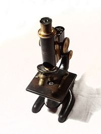
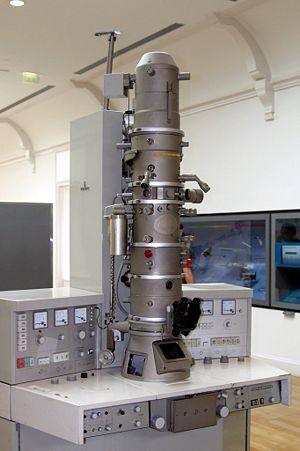
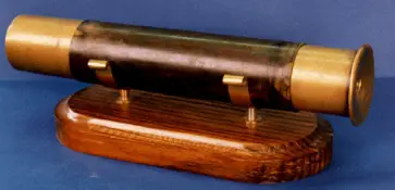
A 1915 Bausch and Lomb microscope
A microscope (Greek: μικρόν (micron) = small + σκοπεῖν (skopein) = to look at) is an instrument for viewing objects that are too small to be seen by the naked or unaided eye. Microscopes give us a larger (magnified) image of a tiny object. The microscopes we use in school and at home trace their history back almost four hundred years.
The most common type of microscope—and the first to be invented—is the optical microscope. This is an optical instrument containing one or more lenses that produce an enlarged image of an object placed in the focal plane of the lens(es). However, human creativity, employed in exploring nature, manifested in numerous improvements and new microscope designs, including the electron microscope, scanning probe microscope, field ion microscope, acoustic microscope, and so forth.
The science of investigating small objects using such an instrument is called microscopy, and the term microscopic means minute or very small, not easily visible with the unaided eye; in other words, requiring a microscope to examine. Microbiology is the scientific study of microorganisms, which are forms of life that are microscopic, such as bacteria, fungi, archaea, and protists.
Microscopes extending the range of human visual sensing reveal a world much grander than the world visible to the unaided eye and remind us of how much we cannot know by our own senses. As new microscopes have revealed new layers of the microscopic world successively further and further removed from the human scale, they have raised new challenges of understanding because objects at different scales have their own distinctive behavior properties, sometimes quite different than those at the human scale. When a scanning tunneling microscope, for example, maps the surface of a crystal and shows us a neat array of fuzzy atoms we can get a better visualization of how the atoms may be arranged, but we also need to remember that those fuzzy balls comprise primarily empty space and that we, ourselves, are no more solid at the atomic level than those fuzzy balls in the picture.
History
The first useful microscope is considered to have been developed in the Netherlands in the late sixteenth and early seventeenth century. There is almost as much confusion about the inventor as about the dates. Three different eyeglass makers have been given credit for the invention: Hans Lippershey (who also developed the first real telescope); Hans Janssen; and his son, Zacharias Janssen. Hans Janssen and Zacharias are often said to have invented the first compound microscope in 1590, but this was a declaration by Zacharias Janssen himself halfway through the seventeenth century. The date is certainly not likely, as it has been shown that Zacharias Janssen actually was born around 1590.
By 1609 Galileo Galilei is also credited with inventing a compound microscope with a convex and a concave lens, which he presented to Polish king Sigismund III in 1612.
In 1625 Giovanni Faber of Bamberg (1574–1629) coined the word microscope by analogy with telescope.
Lens quality in early microscopes was often poor so the images were not very clear. But even these rather crude microscopes were a great help in learning more about animals and plants. For example, in 1665, Robert Hooke (1635–1703) published Micrographia, a collection of biological micrographs developed using the compound microscope. Hooked coined the word cell for the structures he discovered in cork bark.
Thonius Philips van Leeuwenhoek, better known as Anton van Leeuwenhoek (1632–1723), was a Dutch tradesman who is well known for his contribution to the improvement of the microscope, as well as his contributions toward the establishment of microbiology. Known as «the father of microbiology,» Leeuwenhoek, using his handcrafted microscopes, was the first to observe and describe single celled organisms that he first referred to as animalcules, and which we now refer to as microorganisms.
Leeuwenhoek’s interest in microscopes began in 1648 in Amsterdam, when he saw a simple microscope, a magnifying glass mounted on a small stand used by textile merchants capable of magnifying to a power of three. It is believed that soon after 1665, he read Robert Hooke’s book Micrographia, and this roused an interest in using microscopes for the purpose of investigating the natural world.
Leeuwenhoek’s interest in microscopy grew steadily until he was spending most of his nights and free time grinding his own lenses, improving the quality of his microscopes, and studying everything he could beneath them. Although he is sometimes erroneously referred to as «the inventor of the microscope,» compound microscopes (with two lenses mounted together) had existed for about three-quarters of a century. However, they were very crude because the technology used made it difficult to build them properly. Leeuwenhoek’s genius was in developing his skill to grind single lenses very precisely. It is likely that his microscopes were powerful magnifying glasses, not compound microscopes.
During his lifetime Leeuwenhoek ground over five hundred optical lenses. He also created over four hundred different types of microscopes, nine of which still exist today. His microscopes were made of silver or copper metal frames holding hand-ground lenses. Those that survived the years are able to magnify up to 270 times. It is suspected, though, that Leeuwenhoek possessed some microscopes that could magnify up to five hundred times.
In the 1860s, Ernst Abbe discovered the Abbe sine condition, a mathematical condition that must be fulfilled by a lens or other optical system in order for it to produce sharp images of off-axis as well as on-axis objects. This offered a breakthrough in microscope design, which until then was largely based on trial and error. The company of Carl Zeiss exploited this discovery and became the dominant microscope manufacturer of its era.
In 1931 Ernst Ruska started to build the first electron microscope, a transmission electron microscope (TEM). In 1936 Erwin Müller invented the field emission microscope, and in 1951 he invented the field ion microscope and was the first to see atoms. In 1967 he added time-of-flight spectroscopy to the field ion microscope, making the first atom probe and allowing the chemical identification of each individual atom.
In 1953 Frits Zernike, professor of theoretical physics, received the Nobel Prize in Physics for his invention of the phase contrast microscope.
In 1981 Gerd Binnig and Heinrich Rohrer developed the scanning tunneling microscope, and in 1986, Binnig, Quate, and Gerber invented the atomic force microscope. In 1988 Alfred Cerezo, Terence Godfrey, and George Smith applied a position-sensitive detector to the atom probe, making it able to resolve atoms in 3-dimensions. Also in 1988 Kingo Itaya invented the electrochemical scanning tunneling microscope, and in 1991, the Kelvin probe force microscope was invented.
Microscope Types
Microscopes can largely be separated into two classes, optical theory microscopes and scanning probe microscopes.
Optical theory microscopes are microscopes that function through the optical theory of lenses in order to magnify the image generated by the passage of a wave through the sample. The waves used are either electromagnetic in optical microscopes or electron beams in electron microscopes. The types commonly used today are the compound light microscope, stereo microscope, and the electron microscope.
Scanning probe microscopes form images of surfaces using a physical probe that scans the specimen. An image of the surface is obtained by mechanically moving the probe in a raster scan of the specimen, line by line, and recording the probe-surface interaction as a function of position.
Optical microscopes
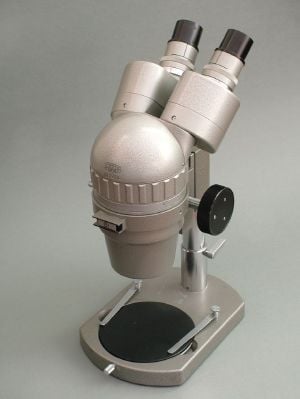
A stereo microscope is often used for lower-power magnification on large subjects
The optical microscope is a type of microscope that uses visible light and a system of lenses to magnify images of small samples. Optical microscopes, through their use of visible wavelengths of light, are the simplest and hence most widely used type of microscope.
There are two basic configurations of optical microscope in use, the simple microscope that uses only one lens for magnification, and the compound microscope that uses a set of many lenses in order to maximize magnification. Recent research [1] has shown that even simple microscopes, those with a single small lens, gave amazingly clear images to the earliest microscopists.
The compound light microscope, in its simplest form—as used by Hooke, for example—would have a single glass lens of short focal length for the objective, and another single glass lens for the eyepiece or ocular. Modern microscopes of this kind are usually more complex, with multiple lens components in both objective and eyepiece assemblies. Today, compound microscopes serve uses in many fields of science, particularly biology and geology.
Optical microscopes use refractive lenses, typically of glass and occasionally of plastic, to focus light into the eye or another light detector. Typical magnification of a light microscope is up to 1,500 times with a theoretical resolution of around 0.2 micrometers. Specialized techniques (e.g., scanning confocal microscopy) may exceed this magnification, but the resolution is an insurmountable diffraction limit.
The stereo or dissecting microscope uses two separate optical paths with two objectives and two eyepieces to provide slightly different viewing angles to the left and right eyes. In this way it produces a three-dimensional (3-D) visualization of the sample being examined. The stereo microscope is often used to study the surfaces of solid specimens or to carry out close work such as sorting, dissection, microsurgery, watch-making, small circuit board manufacture or inspection, and the like.
Electromagnetic wavelengths not visible to human eye
Other microscopes that use electromagnetic wavelengths not visible to the human eye are often called optical microscopes. The most common of these, due to its high resolution yet no requirement for a vacuum-like electron microscope, is the x-ray microscope.
Electron microscopes
Electron microscopes, which use beams of electrons instead of light, are designed for very high magnification usage. Electrons, which have a much smaller wavelength than visible light, allow a much higher resolution. The main limitation of the electron beam is that it must pass through a vacuum as air molecules would otherwise scatter the beam.
Instead of relying on refraction, lenses for electron microscopes are specially designed electromagnets that generates magnetic fields that are approximately parallel to the direction that electrons travel. The electrons are typically detected by a phosphor screen, photographic film or a charge-coupled device (CCD).
Two major variants of electron microscopes exist:
- Scanning electron microscope: looks at the surface of bulk objects by scanning the surface with a fine electron beam and measuring reflection. May also be used for spectroscopy.
- Transmission electron microscope: passes electrons completely through the sample, analogous to basic optical microscopy. This requires careful sample preparation, since electrons are scattered so strongly by most materials. It can also obtain detailed information on the sample’s crystallography through selected area diffraction.
Scanning probe microscope
In scanning probe microscopy (SPM), a physical probe is used either in close contact to the sample or nearly touching it. By rastering the probe across the sample, and by measuring the interactions between the sharp tip of the probe and the sample, a micrograph is generated. The exact nature of the interactions between the probe and the sample determines exactly what kind of SPM is being used. Because this kind of microscopy relies on the interactions between the tip and the sample, it generally only measures information about the surface of the sample.
Some kinds of SPMs are:
- Atomic force microscope
- Scanning tunneling microscope
- Electric force microscope
- Magnetic force microscope (MFM)
- Near-field scanning optical microscope
Point-projection microscopes
The field emission microscope, field ion microscope, and the Atom Probe are examples of point-projection microscopes, where ions are excited from a needle-shaped specimen and hit a detector. The Atom-Probe Tomograph (APT) is the most modern incarnation and allows a three-dimensional atom-by-atom (with chemical elements identified) reconstruction with sub-nanometer resolution.
Acoustic microscopes
Acoustic microscopes use sound waves to measure variations in acoustic impedance. Similar to SONAR in principle, they are used for such jobs as detecting defects in the subsurfaces of materials including those found in integrated circuits.
References
ISBN links support NWE through referral fees
- Dobell, C. (ed.) 1960. Antony van Leeuwenhoek and his «Little Animals. New York: Dover Publications.
- Ford, B. J. 1991. The Leeuwenhoek Legacy. London: Biopress, Bristol, and Farrand Press. ISBN 1850830169.
- Van Berkel, K. “Vermeer, Van Leeuwenhoek en De Astronoom.” Vrij Nederland (February 24, 1996): 62-67.
Credits
New World Encyclopedia writers and editors rewrote and completed the Wikipedia article
in accordance with New World Encyclopedia standards. This article abides by terms of the Creative Commons CC-by-sa 3.0 License (CC-by-sa), which may be used and disseminated with proper attribution. Credit is due under the terms of this license that can reference both the New World Encyclopedia contributors and the selfless volunteer contributors of the Wikimedia Foundation. To cite this article click here for a list of acceptable citing formats.The history of earlier contributions by wikipedians is accessible to researchers here:
- Microscope history
- Timeline_of_microscope_technology history
- Optical_microscope history
The history of this article since it was imported to New World Encyclopedia:
- History of «Microscope»
Note: Some restrictions may apply to use of individual images which are separately licensed.
We have a series of posts about microscope’s function, history, and types.
I. Background and Introduction
- What is a Microscope? – Function and Magnification
- Who Invented the Microscope? – History of Microscopy
- Are Microscopes the Same? – A Introduction of Current Microscope Family
II. Components of a Microscope
III. Specialized Microscopes
IV. Practical Guides to Work with Your Microscopes
The early history of microscopes – from simple glass magnifiers to compound microscopes
A convex lens that can bend and focus light
The key for a microscope to magnify the image is the convex lens.
The oldest objects resembling lenses can date back 3,000 years. This “Nimrud lens” is a crystal disk with a convex shape that belonged to an ancient Near East empire, Assyria. It has been used as a magnifying glass or as a burning-glass to start fires by concentrating sunlight. There were also records showing that ancient Greek know the optical properties of water-filled spheres.
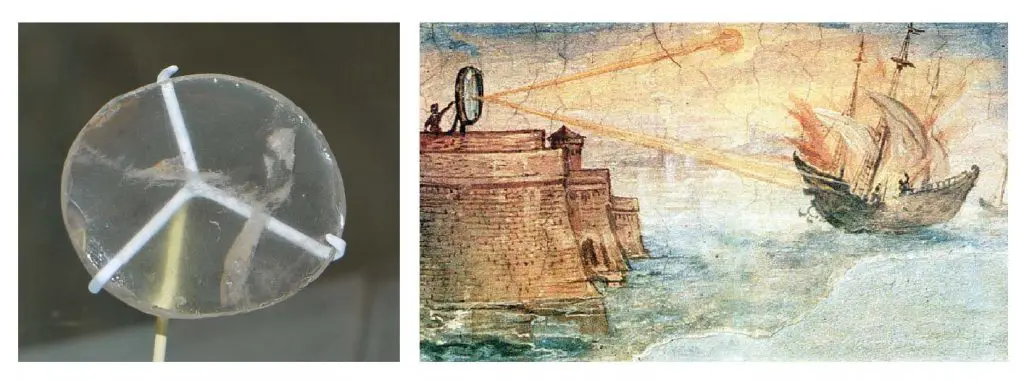
[In this figure] Left: The “Nimrud lens” of Assyrians is a 3000-year-old piece of crystal unearthed in modern-day Iraq. It is displayed in the British Museum, London. Right: A wall painting from the Uffizi Gallery in Florence, Italy, shows the Greek mathematician Archimedes’ mirror burning Roman military ships. Painted in 1600 by Giulio Parigi. Could the mirror really get it hot enough to burst into flame is unknown. However, it proved that ancient Greek may already learn the power of a focused light beam.
First simple microscopes (or magnifying glasses)
The earliest known use of simple microscopes or magnifying glasses (consisted of only one convex lens) dates back to the 13th century. At that time, the use of lenses in eyeglasses became popular in Italy and may lead to the widespread use of simple microscopes. However, these single lens magnifying glasses had only minimal magnification.
Van Leeuwenhoek’s microscopes
Antonie van Leeuwenhoek (1632-1723) was a Dutch businessman and scientist. He is commonly known as “the Father of Microbiology” and one of the first microscopists and microbiologists.
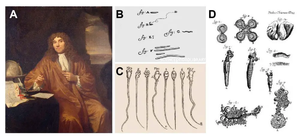
[In this figure] (A) Antony van Leeuwenhoek painted by Jan Verkolje. Van Leeuwenhoek is best known for his pioneering work in microscopy and his contributions toward establishing microbiology as a scientific discipline. (B) Drawings made by Leeuwenhoek of what could be the first bacteria observed. (C) Sperms from rabbit and dog drawn by Leeuwenhoek. (D) Rotifers observed by Leeuwenhoek. He named these tiny creatures “wheel animalcules.”
Antonie van Leeuwenhoek made more than 500 optical lenses. He also created at least 25 single-lens microscopes of different types. Leeuwenhoek modified his microscopes to be capable of magnifying up to 275 times. Using his microscopes, Leeuwenhoek reported the first discovery of protists (he called infusoria) in 1674 and bacteria (he described as “little animals” or animalcules) in 1783. He also observed the vacuole inside the cells, mobility of sperms, and the banded pattern on muscular fibers.
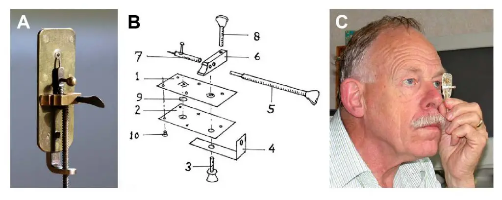
[In this figure] (A) A replica of Van Leeuwenhoek’s microscope. It is only 5-cm long. The lens is in a small hole. The other side of the microscope had a pin, where the sample was attached to stay close to the lens. (B) Exploded design of a van Leeuwenhoek’s microscope. If you want to learn how to make one, check out this great article. (C) The way of using a Van Leeuwenhoek’s microscope. You have to place the lens very close in front of the eye while looking in the direction of the sun. (Don’t look at the sun, it will hurt your eyes.)
[In this video] A cool video showing how to make a paper Leeuwenhoek microscope.
First compound microscopes
The compound microscope’s actual inventor is unknown, although many claims have been made over the years. Among these claims, the Janssen brothers were believed to develop the first compound microscope in the early 1600s.
[In this figure] Janssen’s compound microscope consisted of two convex lenses aligned in series: an object-glass (objective) closer to the object or specimen; and an eyepiece (ocular) closer to the observer’s eye.
Galileo Galilei (Italian astronomer) is also sometimes cited as a compound microscope inventor. He found that he could magnify small objects by viewing through the wrong end of his telescope. After seeing the compound microscope built by others, Galileo built his own improved version.
In 1625, Giovanni Faber coined the name “microscope.” This name came from the Greek words “micro,” meaning “small,” and “scope,” meaning “to look at.”
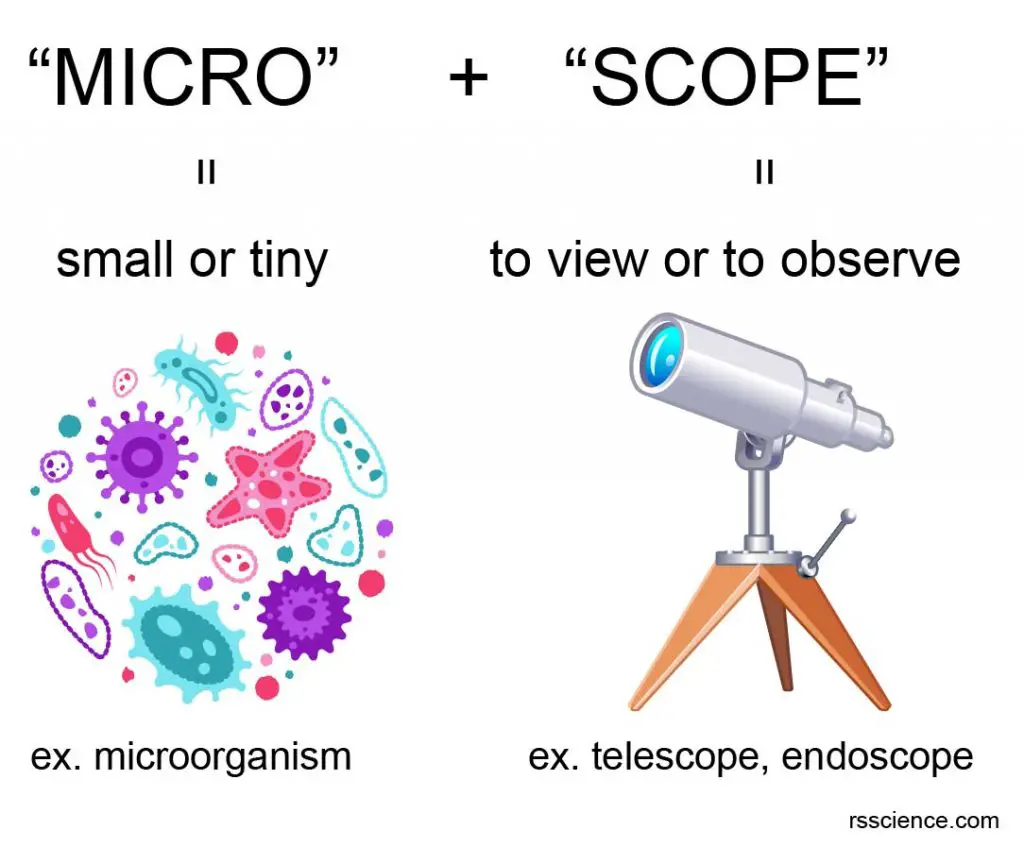
[In this figure] The name “microscope” came from two words – “micro” and “scope”.
Robert Hooke and cell theory
The power of “compound microscopes” allowed Robert Hooke (1635-1703) to establish the cell theory, which states that all organisms are made of cells, all life functions occur in cells, and all cells come from other cells.
While observing cork through his microscope, Hooke saw tiny empty cavities, which he illustrated and described as cells. Hooke’s discovery led to the understanding of cells as the smallest units of life—the foundation of cell theory.
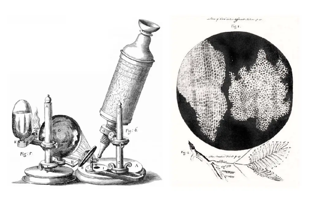
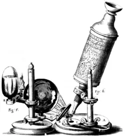
[In this figure] Left: The compound microscope used by Robert Hooke to discover “cells.” Right: Cell structure of cork illuminated by Robert Hooke in Micrographia, 1665.
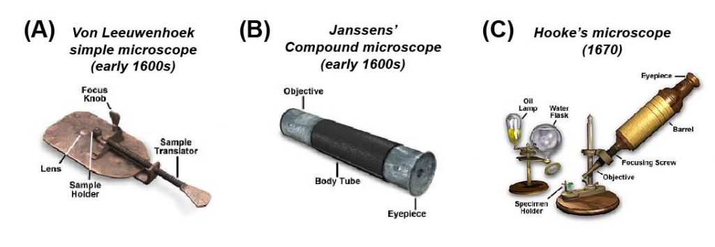
[In this figure] A collection of “antique” microscope dated to the early development of microscopy. (A) Anton von Leeuwenhoek’s microscope was a simple glass magnifier with only one convex lens. He discovered many microorganisms, such as Paramecium, using this simple microscope. (B) Janssen brothers developed the first compound microscope with two aligned convex lenses. It looked very similar to a tubular telescope. (C) Robert Hooke observed “cells” using his modified compound microscope. He used an oil lamp as a light source and a water flask to focus the light beam.
Photo credit: Olympus.
Compound microscopes became a popular and essential scientific instrument
The 18th and 19th centuries witnessed a great improvement in the mechanical and optical quality of compound microscopes. Advances in machine tools allowed more sophisticated optical parts to be fabricated. Newly-invented lenses corrected the optical aberration (both spherical and chromatic) that hampered the quality of microscopic images. Many British and German microscope manufacturers flourished during this time period.
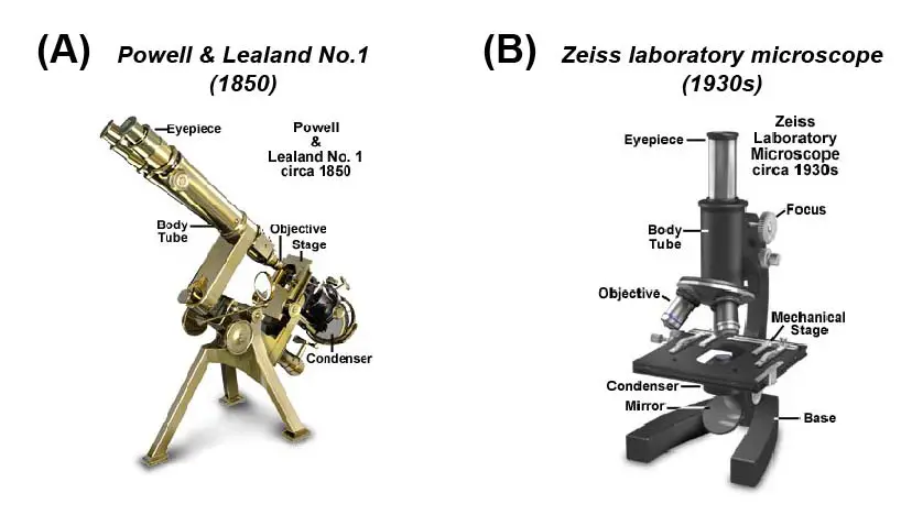
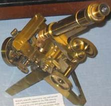
[In this figure] (A) In the 18th and 19th centuries, advances in machine tools allowed more sophisticated microscope parts to be fabricated. Many British and German manufacturers started selling commercial microscopes. The microscope illustrated was manufactured by Hugh Powell and Peter Lealand in London about 1850. (B) The classical design of the Zeiss Laboratory microscope was produced in the early 20th century. This type of microscope is very functional, and many are still in use today.
Photo credit: Olympus.
Light microscopes become a very powerful tool
Today, the family of microscopes was significantly expanded. Microscopes had become an essential instrument in modem technology, research, and industry. In the family tree of an optical (or light) microscope, the principle is still the same: “generating the magnified images through the interaction of visible lights and the subjects.” However, scientists had made tremendous progress to see more specific (such as fluorescence light), deeper (such as the 3D image of cells and tissues), faster (such as automated system), and even smaller (such as beyond the diffraction limit of light). Click the type of microscope to know more.
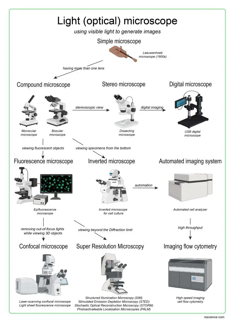
[In this figure] The overview of current light microscopies.
The family tree shows the evolution of light (optical) microscopes.
Electron microscopes change the game
In order to see smaller things, the scientists have to find an illumination with a wavelength much smaller than the visible light. In the early 1930s, they realized that electrons could do so and thus developed the transmission electron microscope (TEM).
According to modern physics, an electron has both properties as a particle and a wave. As a result, an electron has a wavelength (called De Broglie wavelength) depending on its energy (or speed). For example, if an electron is speeded up in a vacuum tube to 1,000,000 meters per second (equal to 2.2 million miles per hour!), the De Broglie wavelength we get is around one-tenth of a nanometer, which is about the size of an atom.
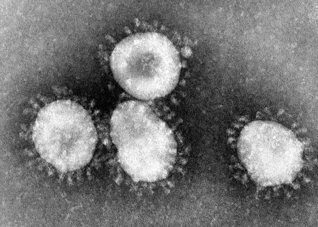
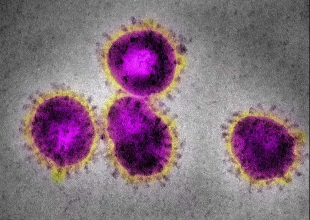
[In this figure] Coronaviruses have a halo or crown-like (corona) appearance.
The new virus, COVID-19, is one kind of coronaviruses. Original electron microscopic images are always black-and-white. The colors are artificially painted for better visualization (right). Photo credit: CDC.
The advance of microscopy never stops
Scientists never give up inventing new types of microscopes and push our limit to see invisible things. For more detail, you can read “the advanced microscopies.”
3D printing techniques also help the development of smaller and cheaper microscopes. You may DIY microscopes (open source) at home and have similar power to professional microscopes in a research laboratory.
Summary
In this article, we learned:
- A simple microscope is the earliest type of microscope. It has only one lens and functions as a magnifying glass.
- Anton von Leeuwenhoek (1632-1723) discovered many microorganisms like protists and bacteria with his simple microscope. He is considered the pioneer of microbiology.
- Compound microscopes use two convex lenses to obtain higher magnification. The first compound microscope was invented in the early 1600s.
- Robert Hooke (1635-1703) observed “cells” using his modified compound microscope and established the cell theory that cells are the basic units of life.
- In the 20th century, microscopes had become the essential instrument and driving force of new technology. Many new microscopes, such as electron microscopy, continue to advance.
References
“Making an Antoni van Leeuwenhoek microscope replica” by Hans Loncke“
“Antony van Leeuwenhoek”
“Anatomy of the Microscope: Introduction”
Olympus