
For a more accessible and less technical introduction to this topic, see Introduction to viruses.
| Virus | |
|---|---|
|
|
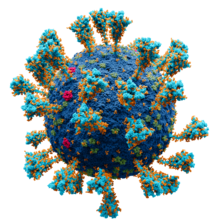
| SARS-CoV-2, a member of the subfamily Coronavirinae | |
| Virus classification |
|
| (unranked): | Virus |
| Realms | |
|
A virus is a submicroscopic infectious agent that replicates only inside the living cells of an organism.[1] Viruses infect all life forms, from animals and plants to microorganisms, including bacteria and archaea.[2][3] Since Dmitri Ivanovsky’s 1892 article describing a non-bacterial pathogen infecting tobacco plants and the discovery of the tobacco mosaic virus by Martinus Beijerinck in 1898,[4] more than 9,000 of the millions of virus species have been described in detail.[5][6] Viruses are found in almost every ecosystem on Earth and are the most numerous type of biological entity.[7][8] The study of viruses is known as virology, a subspeciality of microbiology.
When infected, a host cell is often forced to rapidly produce thousands of copies of the original virus. When not inside an infected cell or in the process of infecting a cell, viruses exist in the form of independent viral particles, or virions, consisting of (i) the genetic material, i.e., long molecules of DNA or RNA that encode the structure of the proteins by which the virus acts; (ii) a protein coat, the capsid, which surrounds and protects the genetic material; and in some cases (iii) an outside envelope of lipids. The shapes of these virus particles range from simple helical and icosahedral forms to more complex structures. Most virus species have virions too small to be seen with an optical microscope and are one-hundredth the size of most bacteria.
The origins of viruses in the evolutionary history of life are unclear: some may have evolved from plasmids—pieces of DNA that can move between cells—while others may have evolved from bacteria. In evolution, viruses are an important means of horizontal gene transfer, which increases genetic diversity in a way analogous to sexual reproduction.[9] Viruses are considered by some biologists to be a life form, because they carry genetic material, reproduce, and evolve through natural selection, although they lack the key characteristics, such as cell structure, that are generally considered necessary criteria for defining life. Because they possess some but not all such qualities, viruses have been described as «organisms at the edge of life»[10] and as replicators.[11]
Viruses spread in many ways. One transmission pathway is through disease-bearing organisms known as vectors: for example, viruses are often transmitted from plant to plant by insects that feed on plant sap, such as aphids; and viruses in animals can be carried by blood-sucking insects. Many viruses, including influenza viruses, SARS-CoV-2, chickenpox, smallpox, and measles, spread in the air by coughing and sneezing. Norovirus and rotavirus, common causes of viral gastroenteritis, are transmitted by the faecal–oral route, passed by hand-to-mouth contact or in food or water. The infectious dose of norovirus required to produce infection in humans is fewer than 100 particles.[12] HIV is one of several viruses transmitted through sexual contact and by exposure to infected blood. The variety of host cells that a virus can infect is called its host range. This can be narrow, meaning a virus is capable of infecting few species, or broad, meaning it is capable of infecting many.[13]
Viral infections in animals provoke an immune response that usually eliminates the infecting virus. Immune responses can also be produced by vaccines, which confer an artificially acquired immunity to the specific viral infection. Some viruses, including those that cause HIV/AIDS, HPV infection, and viral hepatitis, evade these immune responses and result in chronic infections. Several classes of antiviral drugs have been developed.
Etymology
The word is from the Latin neuter vīrus referring to poison and other noxious liquids, from the same Indo-European base as Sanskrit viṣa, Avestan vīša, and ancient Greek ἰός (all meaning ‘poison’), first attested in English in 1398 in John Trevisa’s translation of Bartholomeus Anglicus’s De Proprietatibus Rerum.[14][15] Virulent, from Latin virulentus (‘poisonous’), dates to c. 1400.[16][17] A meaning of ‘agent that causes infectious disease’ is first recorded in 1728,[15] long before the discovery of viruses by Dmitri Ivanovsky in 1892. The English plural is viruses (sometimes also vira),[18] whereas the Latin word is a mass noun, which has no classically attested plural (vīra is used in Neo-Latin[19]). The adjective viral dates to 1948.[20] The term virion (plural virions), which dates from 1959,[21] is also used to refer to a single viral particle that is released from the cell and is capable of infecting other cells of the same type.[22]
Origins
Viruses are found wherever there is life and have probably existed since living cells first evolved.[23] The origin of viruses is unclear because they do not form fossils, so molecular techniques are used to investigate how they arose.[24] In addition, viral genetic material occasionally integrates into the germline of the host organisms, by which they can be passed on vertically to the offspring of the host for many generations. This provides an invaluable source of information for paleovirologists to trace back ancient viruses that have existed up to millions of years ago. There are three main hypotheses that aim to explain the origins of viruses:[25]
- Regressive hypothesis
- Viruses may have once been small cells that parasitised larger cells. Over time, genes not required by their parasitism were lost. The bacteria rickettsia and chlamydia are living cells that, like viruses, can reproduce only inside host cells. They lend support to this hypothesis, as their dependence on parasitism is likely to have caused the loss of genes that enabled them to survive outside a cell. This is also called the ‘degeneracy hypothesis’,[26][27] or ‘reduction hypothesis’.[28]
- Cellular origin hypothesis
- Some viruses may have evolved from bits of DNA or RNA that «escaped» from the genes of a larger organism. The escaped DNA could have come from plasmids (pieces of naked DNA that can move between cells) or transposons (molecules of DNA that replicate and move around to different positions within the genes of the cell).[29] Once called jumping genes, transposons are examples of mobile genetic elements and could be the origin of some viruses. They were discovered in maize by Barbara McClintock in 1950.[30] This is sometimes called the ‘vagrancy hypothesis’,[26][31] or the ‘escape hypothesis’.[28]
- Co-evolution hypothesis
- This is also called the ‘virus-first hypothesis’[28] and proposes that viruses may have evolved from complex molecules of protein and nucleic acid at the same time that cells first appeared on Earth and would have been dependent on cellular life for billions of years. Viroids are molecules of RNA that are not classified as viruses because they lack a protein coat. They have characteristics that are common to several viruses and are often called subviral agents.[32] Viroids are important pathogens of plants.[33] They do not code for proteins but interact with the host cell and use the host machinery for their replication.[34] The hepatitis delta virus of humans has an RNA genome similar to viroids but has a protein coat derived from hepatitis B virus and cannot produce one of its own. It is, therefore, a defective virus. Although hepatitis delta virus genome may replicate independently once inside a host cell, it requires the help of hepatitis B virus to provide a protein coat so that it can be transmitted to new cells.[35] In similar manner, the sputnik virophage is dependent on mimivirus, which infects the protozoan Acanthamoeba castellanii.[36] These viruses, which are dependent on the presence of other virus species in the host cell, are called ‘satellites’ and may represent evolutionary intermediates of viroids and viruses.[37][38]
In the past, there were problems with all of these hypotheses: the regressive hypothesis did not explain why even the smallest of cellular parasites do not resemble viruses in any way. The escape hypothesis did not explain the complex capsids and other structures on virus particles. The virus-first hypothesis contravened the definition of viruses in that they require host cells.[28] Viruses are now recognised as ancient and as having origins that pre-date the divergence of life into the three domains.[39] This discovery has led modern virologists to reconsider and re-evaluate these three classical hypotheses.[39]
The evidence for an ancestral world of RNA cells[40] and computer analysis of viral and host DNA sequences are giving a better understanding of the evolutionary relationships between different viruses and may help identify the ancestors of modern viruses. To date, such analyses have not proved which of these hypotheses is correct.[40] It seems unlikely that all currently known viruses have a common ancestor, and viruses have probably arisen numerous times in the past by one or more mechanisms.[41]
Microbiology
Life properties
Scientific opinions differ on whether viruses are a form of life or organic structures that interact with living organisms.[11] They have been described as «organisms at the edge of life»,[10] since they resemble organisms in that they possess genes, evolve by natural selection,[42] and reproduce by creating multiple copies of themselves through self-assembly. Although they have genes, they do not have a cellular structure, which is often seen as the basic unit of life. Viruses do not have their own metabolism and require a host cell to make new products. They therefore cannot naturally reproduce outside a host cell[43]—although some bacteria such as rickettsia and chlamydia are considered living organisms despite the same limitation.[44][45] Accepted forms of life use cell division to reproduce, whereas viruses spontaneously assemble within cells. They differ from autonomous growth of crystals as they inherit genetic mutations while being subject to natural selection. Virus self-assembly within host cells has implications for the study of the origin of life, as it lends further credence to the hypothesis that life could have started as self-assembling organic molecules.[2]
Structure

Virions of some of the most common human viruses with their relative size. The nucleic acids are not to scale.
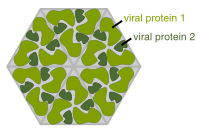
Diagram of how a virus capsid can be constructed using multiple copies of just two protein molecules

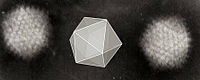
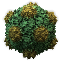
Structure of icosahedral adenovirus. Electron micrograph with an illustration to show shape
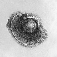
Structure of chickenpox virus. They have a lipid envelope
Viruses display a wide diversity of sizes and shapes, called ‘morphologies’. In general, viruses are much smaller than bacteria and more than a thousand bacteriophage viruses would fit inside an Escherichia coli bacterium’s cell.[46] Many viruses that have been studied are spherical and have a diameter between 20 and 300 nanometres. Some filoviruses, which are filaments, have a total length of up to 1400 nm; their diameters are only about 80 nm.[47] Most viruses cannot be seen with an optical microscope, so scanning and transmission electron microscopes are used to visualise them.[48] To increase the contrast between viruses and the background, electron-dense «stains» are used. These are solutions of salts of heavy metals, such as tungsten, that scatter the electrons from regions covered with the stain. When virions are coated with stain (positive staining), fine detail is obscured. Negative staining overcomes this problem by staining the background only.[49]
A complete virus particle, known as a virion, consists of nucleic acid surrounded by a protective coat of protein called a capsid. These are formed from protein subunits called capsomeres.[50] Viruses can have a lipid «envelope» derived from the host cell membrane. The capsid is made from proteins encoded by the viral genome and its shape serves as the basis for morphological distinction.[51][52] Virally-coded protein subunits will self-assemble to form a capsid, in general requiring the presence of the virus genome. Complex viruses code for proteins that assist in the construction of their capsid. Proteins associated with nucleic acid are known as nucleoproteins, and the association of viral capsid proteins with viral nucleic acid is called a nucleocapsid. The capsid and entire virus structure can be mechanically (physically) probed through atomic force microscopy.[53][54] In general, there are five main morphological virus types:
- Helical
- These viruses are composed of a single type of capsomere stacked around a central axis to form a helical structure, which may have a central cavity, or tube. This arrangement results in virions which can be short and highly rigid rods, or long and very flexible filaments. The genetic material (typically single-stranded RNA, but single-stranded DNA in some cases) is bound into the protein helix by interactions between the negatively charged nucleic acid and positive charges on the protein. Overall, the length of a helical capsid is related to the length of the nucleic acid contained within it, and the diameter is dependent on the size and arrangement of capsomeres. The well-studied tobacco mosaic virus[55] and inovirus[56] are examples of helical viruses.
- Icosahedral
- Most animal viruses are icosahedral or near-spherical with chiral icosahedral symmetry. A regular icosahedron is the optimum way of forming a closed shell from identical subunits. The minimum number of capsomeres required for each triangular face is 3, which gives 60 for the icosahedron. Many viruses, such as rotavirus, have more than 60 capsomers and appear spherical but they retain this symmetry. To achieve this, the capsomeres at the apices are surrounded by five other capsomeres and are called pentons. Capsomeres on the triangular faces are surrounded by six others and are called hexons.[57] Hexons are in essence flat and pentons, which form the 12 vertices, are curved. The same protein may act as the subunit of both the pentamers and hexamers or they may be composed of different proteins.[58]
- Prolate
- This is an icosahedron elongated along the fivefold axis and is a common arrangement of the heads of bacteriophages. This structure is composed of a cylinder with a cap at either end.[59]
- Enveloped
- Some species of virus envelop themselves in a modified form of one of the cell membranes, either the outer membrane surrounding an infected host cell or internal membranes such as a nuclear membrane or endoplasmic reticulum, thus gaining an outer lipid bilayer known as a viral envelope. This membrane is studded with proteins coded for by the viral genome and host genome; the lipid membrane itself and any carbohydrates present originate entirely from the host. Influenza virus, HIV (which causes AIDS), and severe acute respiratory syndrome coronavirus 2 (which causes COVID-19)[60] use this strategy. Most enveloped viruses are dependent on the envelope for their infectivity.[61]
- Complex
- These viruses possess a capsid that is neither purely helical nor purely icosahedral, and that may possess extra structures such as protein tails or a complex outer wall. Some bacteriophages, such as Enterobacteria phage T4, have a complex structure consisting of an icosahedral head bound to a helical tail, which may have a hexagonal base plate with protruding protein tail fibres. This tail structure acts like a molecular syringe, attaching to the bacterial host and then injecting the viral genome into the cell.[62]
The poxviruses are large, complex viruses that have an unusual morphology. The viral genome is associated with proteins within a central disc structure known as a nucleoid. The nucleoid is surrounded by a membrane and two lateral bodies of unknown function. The virus has an outer envelope with a thick layer of protein studded over its surface. The whole virion is slightly pleomorphic, ranging from ovoid to brick-shaped.[63]
Giant viruses
Mimivirus is one of the largest characterised viruses, with a capsid diameter of 400 nm. Protein filaments measuring 100 nm project from the surface. The capsid appears hexagonal under an electron microscope, therefore the capsid is probably icosahedral.[64] In 2011, researchers discovered the largest then known virus in samples of water collected from the ocean floor off the coast of Las Cruces, Chile. Provisionally named Megavirus chilensis, it can be seen with a basic optical microscope.[65] In 2013, the Pandoravirus genus was discovered in Chile and Australia, and has genomes about twice as large as Megavirus and Mimivirus.[66] All giant viruses have dsDNA genomes and they are classified into several families: Mimiviridae, Pithoviridae, Pandoraviridae, Phycodnaviridae, and the Mollivirus genus.[67]
Some viruses that infect Archaea have complex structures unrelated to any other form of virus, with a wide variety of unusual shapes, ranging from spindle-shaped structures to viruses that resemble hooked rods, teardrops or even bottles. Other archaeal viruses resemble the tailed bacteriophages, and can have multiple tail structures.[68]
Genome
| Property | Parameters |
|---|---|
| Nucleic acid |
|
| Shape |
|
| Strandedness |
|
| Sense |
|
An enormous variety of genomic structures can be seen among viral species; as a group, they contain more structural genomic diversity than plants, animals, archaea, or bacteria. There are millions of different types of viruses,[6] although fewer than 7,000 types have been described in detail.[69] As of January 2021, the NCBI Virus genome database has more than 193,000 complete genome sequences,[70] but there are doubtlessly many more to be discovered.[71][72]
A virus has either a DNA or an RNA genome and is called a DNA virus or an RNA virus, respectively. The vast majority of viruses have RNA genomes. Plant viruses tend to have single-stranded RNA genomes and bacteriophages tend to have double-stranded DNA genomes.[73]
Viral genomes are circular, as in the polyomaviruses, or linear, as in the adenoviruses. The type of nucleic acid is irrelevant to the shape of the genome. Among RNA viruses and certain DNA viruses, the genome is often divided up into separate parts, in which case it is called segmented. For RNA viruses, each segment often codes for only one protein and they are usually found together in one capsid. All segments are not required to be in the same virion for the virus to be infectious, as demonstrated by brome mosaic virus and several other plant viruses.[47]
A viral genome, irrespective of nucleic acid type, is almost always either single-stranded (ss) or double-stranded (ds). Single-stranded genomes consist of an unpaired nucleic acid, analogous to one-half of a ladder split down the middle. Double-stranded genomes consist of two complementary paired nucleic acids, analogous to a ladder. The virus particles of some virus families, such as those belonging to the Hepadnaviridae, contain a genome that is partially double-stranded and partially single-stranded.[73]
For most viruses with RNA genomes and some with single-stranded DNA (ssDNA) genomes, the single strands are said to be either positive-sense (called the ‘plus-strand’) or negative-sense (called the ‘minus-strand’), depending on if they are complementary to the viral messenger RNA (mRNA). Positive-sense viral RNA is in the same sense as viral mRNA and thus at least a part of it can be immediately translated by the host cell. Negative-sense viral RNA is complementary to mRNA and thus must be converted to positive-sense RNA by an RNA-dependent RNA polymerase before translation. DNA nomenclature for viruses with genomic ssDNA is similar to RNA nomenclature, in that positive-strand viral ssDNA is identical in sequence to the viral mRNA and is thus a coding strand, while negative-sense viral ssDNA is complementary to the viral mRNA and is thus a template strand.[73] Several types of ssDNA and ssRNA viruses have genomes that are ambisense in that transcription can occur off both strands in a double-stranded replicative intermediate. Examples include geminiviruses, which are ssDNA plant viruses and arenaviruses, which are ssRNA viruses of animals.[74]
Genome size
Genome size varies greatly between species. The smallest—the ssDNA circoviruses, family Circoviridae—code for only two proteins and have a genome size of only two kilobases;[75] the largest—the pandoraviruses—have genome sizes of around two megabases which code for about 2500 proteins.[66] Virus genes rarely have introns and often are arranged in the genome so that they overlap.[76]
In general, RNA viruses have smaller genome sizes than DNA viruses because of a higher error-rate when replicating, and have a maximum upper size limit.[24] Beyond this, errors when replicating render the virus useless or uncompetitive. To compensate, RNA viruses often have segmented genomes—the genome is split into smaller molecules—thus reducing the chance that an error in a single-component genome will incapacitate the entire genome. In contrast, DNA viruses generally have larger genomes because of the high fidelity of their replication enzymes.[77] Single-strand DNA viruses are an exception to this rule, as mutation rates for these genomes can approach the extreme of the ssRNA virus case.[78]
Genetic mutation and recombination

Antigenic shift, or reassortment, can result in novel and highly pathogenic strains of human flu
Viruses undergo genetic change by several mechanisms. These include a process called antigenic drift where individual bases in the DNA or RNA mutate to other bases. Most of these point mutations are «silent»—they do not change the protein that the gene encodes—but others can confer evolutionary advantages such as resistance to antiviral drugs.[79][80] Antigenic shift occurs when there is a major change in the genome of the virus. This can be a result of recombination or reassortment. When this happens with influenza viruses, pandemics might result.[81] RNA viruses often exist as quasispecies or swarms of viruses of the same species but with slightly different genome nucleoside sequences. Such quasispecies are a prime target for natural selection.[82]
Segmented genomes confer evolutionary advantages; different strains of a virus with a segmented genome can shuffle and combine genes and produce progeny viruses (or offspring) that have unique characteristics. This is called reassortment or ‘viral sex’.[83]
Genetic recombination is a process by which a strand of DNA (or RNA) is broken and then joined to the end of a different DNA (or RNA) molecule. This can occur when viruses infect cells simultaneously and studies of viral evolution have shown that recombination has been rampant in the species studied.[84] Recombination is common to both RNA and DNA viruses.[85][86]
Coronaviruses have a single-strand positive-sense RNA genome. Replication of the genome is catalyzed by an RNA-dependent RNA polymerase. The mechanism of recombination used by coronaviruses likely involves template switching by the polymerase during genome replication.[87] This process appears to be an adaptation for coping with genome damage.[88]
Replication cycle
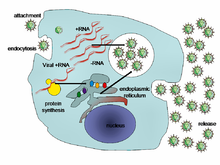
A typical virus replication cycle
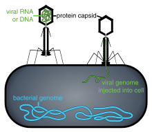
Some bacteriophages inject their genomes into bacterial cells (not to scale)
Viral populations do not grow through cell division, because they are acellular. Instead, they use the machinery and metabolism of a host cell to produce multiple copies of themselves, and they assemble in the cell.[89] When infected, the host cell is forced to rapidly produce thousands of copies of the original virus.[90]
Their life cycle differs greatly between species, but there are six basic stages in their life cycle:[91]
Attachment is a specific binding between viral capsid proteins and specific receptors on the host cellular surface. This specificity determines the host range and type of host cell of a virus. For example, HIV infects a limited range of human leucocytes. This is because its surface protein, gp120, specifically interacts with the CD4 molecule—a chemokine receptor—which is most commonly found on the surface of CD4+ T-Cells. This mechanism has evolved to favour those viruses that infect only cells in which they are capable of replication. Attachment to the receptor can induce the viral envelope protein to undergo changes that result in the fusion of viral and cellular membranes, or changes of non-enveloped virus surface proteins that allow the virus to enter.[92]
Penetration or viral entry follows attachment: Virions enter the host cell through receptor-mediated endocytosis or membrane fusion. The infection of plant and fungal cells is different from that of animal cells. Plants have a rigid cell wall made of cellulose, and fungi one of chitin, so most viruses can get inside these cells only after trauma to the cell wall.[93] Nearly all plant viruses (such as tobacco mosaic virus) can also move directly from cell to cell, in the form of single-stranded nucleoprotein complexes, through pores called plasmodesmata.[94] Bacteria, like plants, have strong cell walls that a virus must breach to infect the cell. Given that bacterial cell walls are much thinner than plant cell walls due to their much smaller size, some viruses have evolved mechanisms that inject their genome into the bacterial cell across the cell wall, while the viral capsid remains outside.[95]
Uncoating is a process in which the viral capsid is removed: This may be by degradation by viral enzymes or host enzymes or by simple dissociation; the end-result is the releasing of the viral genomic nucleic acid.[96]
Replication of viruses involves primarily multiplication of the genome. Replication involves the synthesis of viral messenger RNA (mRNA) from «early» genes (with exceptions for positive-sense RNA viruses), viral protein synthesis, possible assembly of viral proteins, then viral genome replication mediated by early or regulatory protein expression. This may be followed, for complex viruses with larger genomes, by one or more further rounds of mRNA synthesis: «late» gene expression is, in general, of structural or virion proteins.[97]
Assembly – Following the structure-mediated self-assembly of the virus particles, some modification of the proteins often occurs. In viruses such as HIV, this modification (sometimes called maturation) occurs after the virus has been released from the host cell.[98]
Release – Viruses can be released from the host cell by lysis, a process that kills the cell by bursting its membrane and cell wall if present: this is a feature of many bacterial and some animal viruses. Some viruses undergo a lysogenic cycle where the viral genome is incorporated by genetic recombination into a specific place in the host’s chromosome. The viral genome is then known as a «provirus» or, in the case of bacteriophages a «prophage».[99] Whenever the host divides, the viral genome is also replicated. The viral genome is mostly silent within the host. At some point, the provirus or prophage may give rise to the active virus, which may lyse the host cells.[100] Enveloped viruses (e.g., HIV) typically are released from the host cell by budding. During this process, the virus acquires its envelope, which is a modified piece of the host’s plasma or other, internal membrane.[101]
Genome replication
The genetic material within virus particles, and the method by which the material is replicated, varies considerably between different types of viruses.
- DNA viruses
- The genome replication of most DNA viruses takes place in the cell’s nucleus. If the cell has the appropriate receptor on its surface, these viruses enter the cell either by direct fusion with the cell membrane (e.g., herpesviruses) or—more usually—by receptor-mediated endocytosis. Most DNA viruses are entirely dependent on the host cell’s DNA and RNA synthesising machinery and RNA processing machinery. Viruses with larger genomes may encode much of this machinery themselves. In eukaryotes, the viral genome must cross the cell’s nuclear membrane to access this machinery, while in bacteria it need only enter the cell.[102]
- RNA viruses
- Replication of RNA viruses usually takes place in the cytoplasm. RNA viruses can be placed into four different groups depending on their modes of replication. The polarity (whether or not it can be used directly by ribosomes to make proteins) of single-stranded RNA viruses largely determines the replicative mechanism; the other major criterion is whether the genetic material is single-stranded or double-stranded. All RNA viruses use their own RNA replicase enzymes to create copies of their genomes.[103]
- Reverse transcribing viruses
- Reverse transcribing viruses have ssRNA (Retroviridae, Metaviridae, Pseudoviridae) or dsDNA (Caulimoviridae, and Hepadnaviridae) in their particles. Reverse transcribing viruses with RNA genomes (retroviruses) use a DNA intermediate to replicate, whereas those with DNA genomes (pararetroviruses) use an RNA intermediate during genome replication. Both types use a reverse transcriptase, or RNA-dependent DNA polymerase enzyme, to carry out the nucleic acid conversion. Retroviruses integrate the DNA produced by reverse transcription into the host genome as a provirus as a part of the replication process; pararetroviruses do not, although integrated genome copies of especially plant pararetroviruses can give rise to infectious virus.[104] They are susceptible to antiviral drugs that inhibit the reverse transcriptase enzyme, e.g. zidovudine and lamivudine. An example of the first type is HIV, which is a retrovirus. Examples of the second type are the Hepadnaviridae, which includes Hepatitis B virus.[105]
Cytopathic effects on the host cell
The range of structural and biochemical effects that viruses have on the host cell is extensive.[106] These are called ‘cytopathic effects’.[107] Most virus infections eventually result in the death of the host cell. The causes of death include cell lysis, alterations to the cell’s surface membrane and apoptosis.[108] Often cell death is caused by cessation of its normal activities because of suppression by virus-specific proteins, not all of which are components of the virus particle.[109] The distinction between cytopathic and harmless is gradual. Some viruses, such as Epstein–Barr virus, can cause cells to proliferate without causing malignancy,[110] while others, such as papillomaviruses, are established causes of cancer.[111]
Dormant and latent infections
Some viruses cause no apparent changes to the infected cell. Cells in which the virus is latent and inactive show few signs of infection and often function normally.[112] This causes persistent infections and the virus is often dormant for many months or years. This is often the case with herpes viruses.[113][114]
Host range
Viruses are by far the most abundant biological entities on Earth and they outnumber all the others put together.[115] They infect all types of cellular life including animals, plants, bacteria and fungi.[69] Different types of viruses can infect only a limited range of hosts and many are species-specific. Some, such as smallpox virus for example, can infect only one species—in this case humans,[116] and are said to have a narrow host range. Other viruses, such as rabies virus, can infect different species of mammals and are said to have a broad range.[117] The viruses that infect plants are harmless to animals, and most viruses that infect other animals are harmless to humans.[118] The host range of some bacteriophages is limited to a single strain of bacteria and they can be used to trace the source of outbreaks of infections by a method called phage typing.[119] The complete set of viruses in an organism or habitat is called the virome; for example, all human viruses constitute the human virome.[120]
Novel viruses
A novel virus is one that has not previously been recorded. It can be a virus that is isolated from its natural reservoir or isolated as the result of spread to an animal or human host where the virus had not been identified before. It can be an emergent virus, one that represents a new virus, but it can also be an extant virus that has not been previously identified.[121] The SARS-CoV-2 coronavirus that caused the pandemic of covid disease is an example of a novel virus.[122]
Classification
Classification seeks to describe the diversity of viruses by naming and grouping them on the basis of similarities. In 1962, André Lwoff, Robert Horne, and Paul Tournier were the first to develop a means of virus classification, based on the Linnaean hierarchical system.[123] This system based classification on phylum, class, order, family, genus, and species. Viruses were grouped according to their shared properties (not those of their hosts) and the type of nucleic acid forming their genomes.[124] In 1966, the International Committee on Taxonomy of Viruses (ICTV) was formed. The system proposed by Lwoff, Horne and Tournier was initially not accepted by the ICTV because the small genome size of viruses and their high rate of mutation made it difficult to determine their ancestry beyond order. As such, the Baltimore classification system has come to be used to supplement the more traditional hierarchy.[125] Starting in 2018, the ICTV began to acknowledge deeper evolutionary relationships between viruses that have been discovered over time and adopted a 15-rank classification system ranging from realm to species.[126] Additionally, some species within the same genus are grouped into a genogroup.[127][128]
ICTV classification
The ICTV developed the current classification system and wrote guidelines that put a greater weight on certain virus properties to maintain family uniformity. A unified taxonomy (a universal system for classifying viruses) has been established.[129] Only a small part of the total diversity of viruses has been studied.[130] As of 2021, 6 realms, 10 kingdoms, 17 phyla, 2 subphyla, 39 classes, 65 orders, 8 suborders, 233 families, 168 subfamilies, 2,606 genera, 84 subgenera, and 10,434 species of viruses have been defined by the ICTV.[5]
The general taxonomic structure of taxon ranges and the suffixes used in taxonomic names are shown hereafter. As of 2021, the ranks of subrealm, subkingdom, and subclass are unused, whereas all other ranks are in use.[5]
- Realm (-viria)
- Subrealm (-vira)
- Kingdom (-virae)
- Subkingdom (-virites)
- Phylum (-viricota)
- Subphylum (-viricotina)
- Class (-viricetes)
- Subclass (-viricetidae)
- Order (-virales)
- Suborder (-virineae)
- Family (-viridae)
- Subfamily (-virinae)
- Genus (-virus)
- Subgenus (-virus)
- Species
- Subgenus (-virus)
- Genus (-virus)
- Subfamily (-virinae)
- Family (-viridae)
- Suborder (-virineae)
- Order (-virales)
- Subclass (-viricetidae)
- Class (-viricetes)
- Subphylum (-viricotina)
- Phylum (-viricota)
- Subkingdom (-virites)
- Kingdom (-virae)
- Subrealm (-vira)
Baltimore classification

The Baltimore Classification of viruses is based on the method of viral mRNA synthesis
The Nobel Prize-winning biologist David Baltimore devised the Baltimore classification system.[131][132] The ICTV classification system is used in conjunction with the Baltimore classification system in modern virus classification.[133][134][135]
The Baltimore classification of viruses is based on the mechanism of mRNA production. Viruses must generate mRNAs from their genomes to produce proteins and replicate themselves, but different mechanisms are used to achieve this in each virus family. Viral genomes may be single-stranded (ss) or double-stranded (ds), RNA or DNA, and may or may not use reverse transcriptase (RT). In addition, ssRNA viruses may be either sense (+) or antisense (−). This classification places viruses into seven groups:
- I: dsDNA viruses (e.g. Adenoviruses, Herpesviruses, Poxviruses)
- II: ssDNA viruses (+ strand or «sense») DNA (e.g. Parvoviruses)
- III: dsRNA viruses (e.g. Reoviruses)
- IV:(+)ssRNA viruses (+ strand or sense) RNA (e.g. Coronaviruses, Picornaviruses, Togaviruses)
- V: (−)ssRNA viruses (− strand or antisense) RNA (e.g. Orthomyxoviruses, Rhabdoviruses)
- VI: ssRNA-RT viruses (+ strand or sense) RNA with DNA intermediate in life-cycle (e.g. Retroviruses)
- VII: dsDNA-RT viruses DNA with RNA intermediate in life-cycle (e.g. Hepadnaviruses)
Role in human disease

Overview of the main types of viral infection and the most notable species involved[136]
Examples of common human diseases caused by viruses include the common cold, influenza, chickenpox, and cold sores. Many serious diseases such as rabies, Ebola virus disease, AIDS (HIV), avian influenza, and SARS are caused by viruses. The relative ability of viruses to cause disease is described in terms of virulence. Other diseases are under investigation to discover if they have a virus as the causative agent, such as the possible connection between human herpesvirus 6 (HHV6) and neurological diseases such as multiple sclerosis and chronic fatigue syndrome.[137] There is controversy over whether the bornavirus, previously thought to cause neurological diseases in horses, could be responsible for psychiatric illnesses in humans.[138]
Viruses have different mechanisms by which they produce disease in an organism, which depends largely on the viral species. Mechanisms at the cellular level primarily include cell lysis, the breaking open and subsequent death of the cell. In multicellular organisms, if enough cells die, the whole organism will start to suffer the effects. Although viruses cause disruption of healthy homeostasis, resulting in disease, they may exist relatively harmlessly within an organism. An example would include the ability of the herpes simplex virus, which causes cold sores, to remain in a dormant state within the human body. This is called latency[139] and is a characteristic of the herpes viruses, including Epstein–Barr virus, which causes glandular fever, and varicella zoster virus, which causes chickenpox and shingles. Most people have been infected with at least one of these types of herpes virus.[140] These latent viruses might sometimes be beneficial, as the presence of the virus can increase immunity against bacterial pathogens, such as Yersinia pestis.[141]
Some viruses can cause lifelong or chronic infections, where the viruses continue to replicate in the body despite the host’s defence mechanisms.[142] This is common in hepatitis B virus and hepatitis C virus infections. People chronically infected are known as carriers, as they serve as reservoirs of infectious virus.[143] In populations with a high proportion of carriers, the disease is said to be endemic.[144]
Epidemiology
Viral epidemiology is the branch of medical science that deals with the transmission and control of virus infections in humans. Transmission of viruses can be vertical, which means from mother to child, or horizontal, which means from person to person. Examples of vertical transmission include hepatitis B virus and HIV, where the baby is born already infected with the virus.[145] Another, more rare, example is the varicella zoster virus, which, although causing relatively mild infections in children and adults, can be fatal to the foetus and newborn baby.[146]
Horizontal transmission is the most common mechanism of spread of viruses in populations.[147] Horizontal transmission can occur when body fluids are exchanged during sexual activity, by exchange of saliva or when contaminated food or water is ingested. It can also occur when aerosols containing viruses are inhaled or by insect vectors such as when infected mosquitoes penetrate the skin of a host.[147] Most types of viruses are restricted to just one or two of these mechanisms and they are referred to as «respiratory viruses» or «enteric viruses» and so forth. The rate or speed of transmission of viral infections depends on factors that include population density, the number of susceptible individuals, (i.e., those not immune),[148] the quality of healthcare and the weather.[149]
Epidemiology is used to break the chain of infection in populations during outbreaks of viral diseases.[150] Control measures are used that are based on knowledge of how the virus is transmitted. It is important to find the source, or sources, of the outbreak and to identify the virus. Once the virus has been identified, the chain of transmission can sometimes be broken by vaccines. When vaccines are not available, sanitation and disinfection can be effective. Often, infected people are isolated from the rest of the community, and those that have been exposed to the virus are placed in quarantine.[151] To control the outbreak of foot-and-mouth disease in cattle in Britain in 2001, thousands of cattle were slaughtered.[152] Most viral infections of humans and other animals have incubation periods during which the infection causes no signs or symptoms.[153] Incubation periods for viral diseases range from a few days to weeks, but are known for most infections.[154] Somewhat overlapping, but mainly following the incubation period, there is a period of communicability—a time when an infected individual or animal is contagious and can infect another person or animal.[154] This, too, is known for many viral infections, and knowledge of the length of both periods is important in the control of outbreaks.[155] When outbreaks cause an unusually high proportion of cases in a population, community, or region, they are called epidemics. If outbreaks spread worldwide, they are called pandemics.[156]
Epidemics and pandemics

A pandemic is a worldwide epidemic. The 1918 flu pandemic, which lasted until 1919, was a category 5 influenza pandemic caused by an unusually severe and deadly influenza A virus. The victims were often healthy young adults, in contrast to most influenza outbreaks, which predominantly affect juvenile, elderly, or otherwise-weakened patients.[157] Older estimates say it killed 40–50 million people,[158] while more recent research suggests that it may have killed as many as 100 million people, or 5% of the world’s population in 1918.[159]
Although viral pandemics are rare events, HIV—which evolved from viruses found in monkeys and chimpanzees—has been pandemic since at least the 1980s.[160] During the 20th century there were four pandemics caused by influenza virus and those that occurred in 1918, 1957 and 1968 were severe.[161] Most researchers believe that HIV originated in sub-Saharan Africa during the 20th century;[162] it is now a pandemic, with an estimated 37.9 million people now living with the disease worldwide.[163] There were about 770,000 deaths from AIDS in 2018.[164] The Joint United Nations Programme on HIV/AIDS (UNAIDS) and the World Health Organization (WHO) estimate that AIDS has killed more than 25 million people since it was first recognised on 5 June 1981, making it one of the most destructive epidemics in recorded history.[165] In 2007 there were 2.7 million new HIV infections and 2 million HIV-related deaths.[166]


Ebola (top) and Marburg viruses (bottom)
Several highly lethal viral pathogens are members of the Filoviridae. Filoviruses are filament-like viruses that cause viral hemorrhagic fever, and include ebolaviruses and marburgviruses. Marburg virus, first discovered in 1967, attracted widespread press attention in April 2005 for an outbreak in Angola.[167] Ebola virus disease has also caused intermittent outbreaks with high mortality rates since 1976 when it was first identified. The worst and most recent one is the 2013–2016 West Africa epidemic.[168]
Except for smallpox, most pandemics are caused by newly evolved viruses. These «emergent» viruses are usually mutants of less harmful viruses that have circulated previously either in humans or other animals.[169]
Severe acute respiratory syndrome (SARS) and Middle East respiratory syndrome (MERS) are caused by new types of coronaviruses. Other coronaviruses are known to cause mild infections in humans,[170] so the virulence and rapid spread of SARS infections—that by July 2003 had caused around 8,000 cases and 800 deaths—was unexpected and most countries were not prepared.[171]
A related coronavirus, severe acute respiratory syndrome coronavirus 2 (SARS-Cov-2), thought to have originated in bats, emerged in Wuhan, China in November 2019 and spread rapidly around the world. Infections with the virus caused the COVID-19 pandemic that started in 2020.[122][172][173] Unprecedented restrictions in peacetime were placed on international travel,[174] and curfews were imposed in several major cities worldwide in response to the pandemic.[175]
Cancer
Viruses are an established cause of cancer in humans and other species. Viral cancers occur only in a minority of infected persons (or animals). Cancer viruses come from a range of virus families, including both RNA and DNA viruses, and so there is no single type of «oncovirus» (an obsolete term originally used for acutely transforming retroviruses). The development of cancer is determined by a variety of factors such as host immunity[176] and mutations in the host.[177] Viruses accepted to cause human cancers include some genotypes of human papillomavirus, hepatitis B virus, hepatitis C virus, Epstein–Barr virus, Kaposi’s sarcoma-associated herpesvirus and human T-lymphotropic virus. The most recently discovered human cancer virus is a polyomavirus (Merkel cell polyomavirus) that causes most cases of a rare form of skin cancer called Merkel cell carcinoma.[178]
Hepatitis viruses can develop into a chronic viral infection that leads to liver cancer.[179][180] Infection by human T-lymphotropic virus can lead to tropical spastic paraparesis and adult T-cell leukaemia.[181] Human papillomaviruses are an established cause of cancers of cervix, skin, anus, and penis.[182] Within the Herpesviridae, Kaposi’s sarcoma-associated herpesvirus causes Kaposi’s sarcoma and body-cavity lymphoma, and Epstein–Barr virus causes Burkitt’s lymphoma, Hodgkin’s lymphoma, B lymphoproliferative disorder, and nasopharyngeal carcinoma.[183] Merkel cell polyomavirus closely related to SV40 and mouse polyomaviruses that have been used as animal models for cancer viruses for over 50 years.[184]
Host defence mechanisms
The body’s first line of defence against viruses is the innate immune system. This comprises cells and other mechanisms that defend the host from infection in a non-specific manner. This means that the cells of the innate system recognise, and respond to, pathogens in a generic way, but, unlike the adaptive immune system, it does not confer long-lasting or protective immunity to the host.[185]
RNA interference is an important innate defence against viruses.[186] Many viruses have a replication strategy that involves double-stranded RNA (dsRNA). When such a virus infects a cell, it releases its RNA molecule or molecules, which immediately bind to a protein complex called a dicer that cuts the RNA into smaller pieces. A biochemical pathway—the RISC complex—is activated, which ensures cell survival by degrading the viral mRNA. Rotaviruses have evolved to avoid this defence mechanism by not uncoating fully inside the cell, and releasing newly produced mRNA through pores in the particle’s inner capsid. Their genomic dsRNA remains protected inside the core of the virion.[187][188]
When the adaptive immune system of a vertebrate encounters a virus, it produces specific antibodies that bind to the virus and often render it non-infectious. This is called humoral immunity. Two types of antibodies are important. The first, called IgM, is highly effective at neutralising viruses but is produced by the cells of the immune system only for a few weeks. The second, called IgG, is produced indefinitely. The presence of IgM in the blood of the host is used to test for acute infection, whereas IgG indicates an infection sometime in the past.[189] IgG antibody is measured when tests for immunity are carried out.[190]
Antibodies can continue to be an effective defence mechanism even after viruses have managed to gain entry to the host cell. A protein that is in cells, called TRIM21, can attach to the antibodies on the surface of the virus particle. This primes the subsequent destruction of the virus by the enzymes of the cell’s proteosome system.[191]

Two rotaviruses: the one on the right is coated with antibodies that prevent its attachment to cells and infecting them.
A second defence of vertebrates against viruses is called cell-mediated immunity and involves immune cells known as T cells. The body’s cells constantly display short fragments of their proteins on the cell’s surface, and, if a T cell recognises a suspicious viral fragment there, the host cell is destroyed by ‘killer T’ cells and the virus-specific T-cells proliferate. Cells such as the macrophage are specialists at this antigen presentation.[192] The production of interferon is an important host defence mechanism. This is a hormone produced by the body when viruses are present. Its role in immunity is complex; it eventually stops the viruses from reproducing by killing the infected cell and its close neighbours.[193]
Not all virus infections produce a protective immune response in this way. HIV evades the immune system by constantly changing the amino acid sequence of the proteins on the surface of the virion. This is known as «escape mutation» as the viral epitopes escape recognition by the host immune response. These persistent viruses evade immune control by sequestration, blockade of antigen presentation, cytokine resistance, evasion of natural killer cell activities, escape from apoptosis, and antigenic shift.[194] Other viruses, called ‘neurotropic viruses’, are disseminated by neural spread where the immune system may be unable to reach them due to immune privilege.[195]
Prevention and treatment
Because viruses use vital metabolic pathways within host cells to replicate, they are difficult to eliminate without using drugs that cause toxic effects to host cells in general. The most effective medical approaches to viral diseases are vaccinations to provide immunity to infection, and antiviral drugs that selectively interfere with viral replication.
Vaccines
Vaccination is a cheap and effective way of preventing infections by viruses. Vaccines were used to prevent viral infections long before the discovery of the actual viruses. Their use has resulted in a dramatic decline in morbidity (illness) and mortality (death) associated with viral infections such as polio, measles, mumps and rubella.[196] Smallpox infections have been eradicated.[197] Vaccines are available to prevent over thirteen viral infections of humans,[198] and more are used to prevent viral infections of animals.[199] Vaccines can consist of live-attenuated or killed viruses, viral proteins (antigens), or RNA.[200][201] Live vaccines contain weakened forms of the virus, which do not cause the disease but, nonetheless, confer immunity. Such viruses are called attenuated. Live vaccines can be dangerous when given to people with a weak immunity (who are described as immunocompromised), because in these people, the weakened virus can cause the original disease.[202] Biotechnology and genetic engineering techniques are used to produce subunit vaccines. These vaccines use only the capsid proteins of the virus. Hepatitis B vaccine is an example of this type of vaccine.[203] Subunit vaccines are safe for immunocompromised patients because they cannot cause the disease.[204] The yellow fever virus vaccine, a live-attenuated strain called 17D, is probably the safest and most effective vaccine ever generated.[205]
Antiviral drugs

Antiviral drugs are often nucleoside analogues (fake DNA building-blocks), which viruses mistakenly incorporate into their genomes during replication.[206] The life-cycle of the virus is then halted because the newly synthesised DNA is inactive. This is because these analogues lack the hydroxyl groups, which, along with phosphorus atoms, link together to form the strong «backbone» of the DNA molecule. This is called DNA chain termination.[207] Examples of nucleoside analogues are aciclovir for Herpes simplex virus infections and lamivudine for HIV and hepatitis B virus infections. Aciclovir is one of the oldest and most frequently prescribed antiviral drugs.[208]
Other antiviral drugs in use target different stages of the viral life cycle. HIV is dependent on a proteolytic enzyme called the HIV-1 protease for it to become fully infectious. There is a large class of drugs called protease inhibitors that inactivate this enzyme.[209] There are around thirteen classes of antiviral drugs each targeting different viruses or stages of viral replication.[206]
Hepatitis C is caused by an RNA virus. In 80% of people infected, the disease is chronic, and without treatment, they are infected for the remainder of their lives. There are effective treatments that use direct-acting antivirals.[210] The treatment of chronic carriers of the hepatitis B virus has also been developed by using similar strategies that include lamivudine and other anti-viral drugs.[211]
Infection in other species
Viruses infect all cellular life and, although viruses occur universally, each cellular species has its own specific range that often infects only that species.[212] Some viruses, called satellites, can replicate only within cells that have already been infected by another virus.[36]
Animal viruses
Viruses are important pathogens of livestock. Diseases such as foot-and-mouth disease and bluetongue are caused by viruses.[213] Companion animals such as cats, dogs, and horses, if not vaccinated, are susceptible to serious viral infections. Canine parvovirus is caused by a small DNA virus and infections are often fatal in pups.[214] Like all invertebrates, the honey bee is susceptible to many viral infections.[215] Most viruses co-exist harmlessly in their host and cause no signs or symptoms of disease.[4]
Plant viruses

Peppers infected by mild mottle virus
There are many types of plant viruses, but often they cause only a loss of yield, and it is not economically viable to try to control them. Plant viruses are often spread from plant to plant by organisms, known as vectors. These are usually insects, but some fungi, nematode worms, single-celled organisms, and parasitic plants are vectors.[216] When control of plant virus infections is considered economical, for perennial fruits, for example, efforts are concentrated on killing the vectors and removing alternate hosts such as weeds.[217] Plant viruses cannot infect humans and other animals because they can reproduce only in living plant cells.[218]
Originally from Peru, the potato has become a staple crop worldwide.[219] The potato virus Y causes disease in potatoes and related species including tomatoes and peppers. In the 1980s, this virus acquired economical importance when it proved difficult to control in seed potato crops. Transmitted by aphids, this virus can reduce crop yields by up to 80 per cent, causing significant losses to potato yields.[220]
Plants have elaborate and effective defence mechanisms against viruses. One of the most effective is the presence of so-called resistance (R) genes. Each R gene confers resistance to a particular virus by triggering localised areas of cell death around the infected cell, which can often be seen with the unaided eye as large spots. This stops the infection from spreading.[221] RNA interference is also an effective defence in plants.[222] When they are infected, plants often produce natural disinfectants that kill viruses, such as salicylic acid, nitric oxide, and reactive oxygen molecules.[223]
Plant virus particles or virus-like particles (VLPs) have applications in both biotechnology and nanotechnology. The capsids of most plant viruses are simple and robust structures and can be produced in large quantities either by the infection of plants or by expression in a variety of heterologous systems. Plant virus particles can be modified genetically and chemically to encapsulate foreign material and can be incorporated into supramolecular structures for use in biotechnology.[224]
Bacterial viruses

Transmission electron micrograph of multiple bacteriophages attached to a bacterial cell wall
Bacteriophages are a common and diverse group of viruses and are the most abundant biological entity in aquatic environments—there are up to ten times more of these viruses in the oceans than there are bacteria,[225] reaching levels of 250,000,000 bacteriophages per millilitre of seawater.[226] These viruses infect specific bacteria by binding to surface receptor molecules and then entering the cell. Within a short amount of time, in some cases, just minutes, bacterial polymerase starts translating viral mRNA into protein. These proteins go on to become either new virions within the cell, helper proteins, which help assembly of new virions, or proteins involved in cell lysis. Viral enzymes aid in the breakdown of the cell membrane, and, in the case of the T4 phage, in just over twenty minutes after injection over three hundred phages could be released.[227]
The major way bacteria defend themselves from bacteriophages is by producing enzymes that destroy foreign DNA. These enzymes, called restriction endonucleases, cut up the viral DNA that bacteriophages inject into bacterial cells.[228] Bacteria also contain a system that uses CRISPR sequences to retain fragments of the genomes of viruses that the bacteria have come into contact with in the past, which allows them to block the virus’s replication through a form of RNA interference.[229][230] This genetic system provides bacteria with acquired immunity to infection.[231]
Archaeal viruses
Some viruses replicate within archaea: these are DNA viruses with unusual and sometimes unique shapes.[7][68] These viruses have been studied in most detail in the thermophilic archaea, particularly the orders Sulfolobales and Thermoproteales.[232] Defences against these viruses involve RNA interference from repetitive DNA sequences within archaean genomes that are related to the genes of the viruses.[233][234] Most archaea have CRISPR–Cas systems as an adaptive defence against viruses. These enable archaea to retain sections of viral DNA, which are then used to target and eliminate subsequent infections by the virus using a process similar to RNA interference.[235]
Role in aquatic ecosystems
Viruses are the most abundant biological entity in aquatic environments.[2] There are about ten million of them in a teaspoon of seawater.[236] Most of these viruses are bacteriophages infecting heterotrophic bacteria and cyanophages infecting cyanobacteria and they are essential to the regulation of saltwater and freshwater ecosystems.[237]
Bacteriophages are harmless to plants and animals, and are essential to the regulation of marine and freshwater ecosystems[238] are important mortality agents of phytoplankton, the base of the foodchain in aquatic environments.[239] They infect and destroy bacteria in aquatic microbial communities, and are one of the most important mechanisms of recycling carbon and nutrient cycling in marine environments. The organic molecules released from the dead bacterial cells stimulate fresh bacterial and algal growth, in a process known as the viral shunt.[240] In particular, lysis of bacteria by viruses has been shown to enhance nitrogen cycling and stimulate phytoplankton growth.[241] Viral activity may also affect the biological pump, the process whereby carbon is sequestered in the deep ocean.[242]
Microorganisms constitute more than 90% of the biomass in the sea. It is estimated that viruses kill approximately 20% of this biomass each day and that there are 10 to 15 times as many viruses in the oceans as there are bacteria and archaea.[243] Viruses are also major agents responsible for the destruction of phytoplankton including harmful algal blooms,[244]
The number of viruses in the oceans decreases further offshore and deeper into the water, where there are fewer host organisms.[242]
In January 2018, scientists reported that 800 million viruses, mainly of marine origin, are deposited daily from the Earth‘s atmosphere onto every square meter of the planet’s surface, as the result of a global atmospheric stream of viruses, circulating above the weather system but below the altitude of usual airline travel, distributing viruses around the planet.[245][246]
Like any organism, marine mammals are susceptible to viral infections. In 1988 and 2002, thousands of harbour seals were killed in Europe by phocine distemper virus.[247] Many other viruses, including caliciviruses, herpesviruses, adenoviruses and parvoviruses, circulate in marine mammal populations.[242]
In December 2022, scientists reported the first observation of virovory via an experiment on pond water containing chlorovirus, which commonly infects green algae in freshwater environments. When all other microbial food sources were removed from the water, the ciliate Halteria was observed to have increased in number due to the active consumption of chlorovirus as a food source instead of its typical bacterivore diet.[248][249]
Role in evolution
Viruses are an important natural means of transferring genes between different species, which increases genetic diversity and drives evolution.[9][250] It is thought that viruses played a central role in early evolution, before the diversification of the last universal common ancestor into bacteria, archaea and eukaryotes.[251] Viruses are still one of the largest reservoirs of unexplored genetic diversity on Earth.[242]
Applications
Life sciences and medicine

Scientist studying the H5N1 influenza virus
Viruses are important to the study of molecular and cell biology as they provide simple systems that can be used to manipulate and investigate the functions of cells.[252] The study and use of viruses have provided valuable information about aspects of cell biology.[253] For example, viruses have been useful in the study of genetics and helped our understanding of the basic mechanisms of molecular genetics, such as DNA replication, transcription, RNA processing, translation, protein transport, and immunology.
Geneticists often use viruses as vectors to introduce genes into cells that they are studying. This is useful for making the cell produce a foreign substance, or to study the effect of introducing a new gene into the genome. Similarly, virotherapy uses viruses as vectors to treat various diseases, as they can specifically target cells and DNA. It shows promising use in the treatment of cancer and in gene therapy. Eastern European scientists have used phage therapy as an alternative to antibiotics for some time, and interest in this approach is increasing, because of the high level of antibiotic resistance now found in some pathogenic bacteria.[254]
The expression of heterologous proteins by viruses is the basis of several manufacturing processes that are currently being used for the production of various proteins such as vaccine antigens and antibodies. Industrial processes have been recently developed using viral vectors and several pharmaceutical proteins are currently in pre-clinical and clinical trials.[255]
Virotherapy
Virotherapy involves the use of genetically modified viruses to treat diseases.[256] Viruses have been modified by scientists to reproduce in cancer cells and destroy them but not infect healthy cells. Talimogene laherparepvec (T-VEC), for example, is a modified herpes simplex virus that has had a gene, which is required for viruses to replicate in healthy cells, deleted and replaced with a human gene (GM-CSF) that stimulates immunity. When this virus infects cancer cells, it destroys them and in doing so the presence the GM-CSF gene attracts dendritic cells from the surrounding tissues of the body. The dendritic cells process the dead cancer cells and present components of them to other cells of the immune system.[257] Having completed successful clinical trials, the virus gained approval for the treatment of melanoma in late 2015.[258] Viruses that have been reprogrammed to kill cancer cells are called oncolytic viruses.[259]
Materials science and nanotechnology
Current trends in nanotechnology promise to make much more versatile use of viruses.[260] From the viewpoint of a materials scientist, viruses can be regarded as organic nanoparticles.
Their surface carries specific tools that enable them to cross the barriers of their host cells. The size and shape of viruses and the number and nature of the functional groups on their surface are precisely defined. As such, viruses are commonly used in materials science as scaffolds for covalently linked surface modifications. A particular quality of viruses is that they can be tailored by directed evolution. The powerful techniques developed by life sciences are becoming the basis of engineering approaches towards nanomaterials, opening a wide range of applications far beyond biology and medicine.[261]
Because of their size, shape, and well-defined chemical structures, viruses have been used as templates for organising materials on the nanoscale. Recent examples include work at the Naval Research Laboratory in Washington, D.C., using Cowpea mosaic virus (CPMV) particles to amplify signals in DNA microarray based sensors. In this application, the virus particles separate the fluorescent dyes used for signalling to prevent the formation of non-fluorescent dimers that act as quenchers.[262] Another example is the use of CPMV as a nanoscale breadboard for molecular electronics.[263]
Synthetic viruses
Many viruses can be synthesised de novo («from scratch»). The first synthetic virus was created in 2002.[264] Although somewhat of a misconception, it is not the actual virus that is synthesised, but rather its DNA genome (in case of a DNA virus), or a cDNA copy of its genome (in case of RNA viruses). For many virus families the naked synthetic DNA or RNA (once enzymatically converted back from the synthetic cDNA) is infectious when introduced into a cell. That is, they contain all the necessary information to produce new viruses. This technology is now being used to investigate novel vaccine strategies.[265] The ability to synthesise viruses has far-reaching consequences, since viruses can no longer be regarded as extinct, as long as the information of their genome sequence is known and permissive cells are available. As of June 2021, the full-length genome sequences of 11,464 different viruses, including smallpox, are publicly available in an online database maintained by the National Institutes of Health.[266]
Weapons
The ability of viruses to cause devastating epidemics in human societies has led to the concern that viruses could be weaponised for biological warfare. Further concern was raised by the successful recreation of the infamous 1918 influenza virus in a laboratory.[267]
The smallpox virus devastated numerous societies throughout history before its eradication. There are only two centres in the world authorised by the WHO to keep stocks of smallpox virus: the State Research Center of Virology and Biotechnology VECTOR in Russia and the Centers for Disease Control and Prevention in the United States.[268] It may be used as a weapon,[268] as the vaccine for smallpox sometimes had severe side-effects, it is no longer used routinely in any country. Thus, much of the modern human population has almost no established resistance to smallpox and would be vulnerable to the virus.[268]
See also
- Cross-species transmission
- Glossary of virology
- Law of declining virulence – Disproved hypothesis of epidemiologist Theobald Smith
- Non-cellular life
- Retrozyme
- Theory of virulence – Theory by biologist Paul W. Ewald
- Viral metagenomics
- Viroplasm
- Zoonosis
References
Notes
- ^ Wu KJ (15 April 2020). «There are more viruses than stars in the universe. Why do only some infect us? – More than a quadrillion quadrillion individual viruses exist on Earth, but most are not poised to hop into humans. Can we find the ones that are?». National Geographic Society. Retrieved 18 May 2020.
- ^ a b c Koonin EV, Senkevich TG, Dolja VV (September 2006). «The ancient Virus World and evolution of cells». Biology Direct. 1 (1): 29. doi:10.1186/1745-6150-1-29. PMC 1594570. PMID 16984643.
- ^ Zimmer C (26 February 2021). «The Secret Life of a Coronavirus — An oily, 100-nanometer-wide bubble of genes has killed more than two million people and reshaped the world. Scientists don’t quite know what to make of it». The New York Times. Archived from the original on 28 December 2021. Retrieved 28 February 2021.
- ^ a b Dimmock p. 4
- ^ a b c «Virus Taxonomy: 2021 Release». talk.ictvonline.org. International Committee on Taxonomy of Viruses. Retrieved 4 April 2022.
- ^ a b Breitbart M, Rohwer F (June 2005). «Here a virus, there a virus, everywhere the same virus?». Trends in Microbiology. 13 (6): 278–84. doi:10.1016/j.tim.2005.04.003. PMID 15936660.
- ^ a b Lawrence CM, Menon S, Eilers BJ, Bothner B, Khayat R, Douglas T, Young MJ (May 2009). «Structural and functional studies of archaeal viruses». The Journal of Biological Chemistry. 284 (19): 12599–603. doi:10.1074/jbc.R800078200. PMC 2675988. PMID 19158076.
- ^ Edwards RA, Rohwer F (June 2005). «Viral metagenomics». Nature Reviews. Microbiology. 3 (6): 504–10. doi:10.1038/nrmicro1163. PMID 15886693. S2CID 8059643.
- ^ a b Canchaya C, Fournous G, Chibani-Chennoufi S, Dillmann ML, Brüssow H (August 2003). «Phage as agents of lateral gene transfer». Current Opinion in Microbiology. 6 (4): 417–24. doi:10.1016/S1369-5274(03)00086-9. PMID 12941415.
- ^ a b Rybicki EP (1990). «The classification of organisms at the edge of life, or problems with virus systematics». South African Journal of Science. 86: 182–86.
- ^ a b Koonin EV, Starokadomskyy P (October 2016). «Are viruses alive? The replicator paradigm sheds decisive light on an old but misguided question». Studies in History and Philosophy of Biological and Biomedical Sciences. 59: 125–34. doi:10.1016/j.shpsc.2016.02.016. PMC 5406846. PMID 26965225.
- ^ Robilotti E, Deresinski S, Pinsky BA (January 2015). «Norovirus». Clinical Microbiology Reviews. 28 (1): 134–64. doi:10.1128/CMR.00075-14. PMC 4284304. PMID 25567225.
- ^ Shors pp. 123–124
- ^ «Virus, n.». OED Online. Oxford University Press. March 2015.
- ^ a b Harper D (2011). «virus». The Online Etymology Dictionary. Retrieved 19 December 2014.
- ^ «Virulent, adj.». OED Online. Oxford University Press. March 2015.
- ^ Harper D (2011). «virulent». The Online Etymology Dictionary. Retrieved 19 December 2014.
- ^ Buschard K, Thon R (2003). «Diabetic Animal Models». In Hau J, Van Hoosier Jr GL (eds.). Handbook of Laboratory Animal Science. Animal Models. Vol. II (Second ed.). CRC Press. pp. 163, 166.
- ^ William T. Stearn: Botanical Latin. History, Grammar, Syntax, Terminology and Vocabulary. David & Charles, third edition, 1983. Quote: «Virus: virus (s.n. II), gen. sing. viri, nom. pl. vira, gen. pl. vīrorum (to be distinguished from virorum, of men).»
- ^ Harper D (2011). «viral». The Online Etymology Dictionary. Retrieved 19 December 2014.
- ^ Harper D (2011). «virion». The Online Etymology Dictionary. Retrieved 19 December 2014.
- ^ Casjens S (2010). Mahy BW, Van Regenmortel MH (eds.). Desk Encyclopedia of General Virology. Boston: Academic Press. p. 167. ISBN 978-0-12-375146-1.
- ^ Iyer LM, Balaji S, Koonin EV, Aravind L (April 2006). «Evolutionary genomics of nucleo-cytoplasmic large DNA viruses». Virus Research. 117 (1): 156–84. doi:10.1016/j.virusres.2006.01.009. PMID 16494962.
- ^ a b Sanjuán R, Nebot MR, Chirico N, Mansky LM, Belshaw R (October 2010). «Viral mutation rates». Journal of Virology. 84 (19): 9733–48. doi:10.1128/JVI.00694-10. PMC 2937809. PMID 20660197.
- ^ Krupovic M, Dolja VV, Koonin EV (July 2019). «Origin of viruses: primordial replicators recruiting capsids from hosts» (PDF). Nature Reviews. Microbiology. 17 (7): 449–458. doi:10.1038/s41579-019-0205-6. PMID 31142823. S2CID 256744818.
- ^ a b Dimmock p. 16
- ^ Collier p. 11
- ^ a b c d Mahy WJ, Regenmortel MH, eds. (2009). Desk Encyclopedia of General Virology. Oxford: Academic Press. p. 24. ISBN 978-0-12-375146-1.
- ^ Shors p. 810
- ^ McClintock B (June 1950). «The origin and behavior of mutable loci in maize». Proceedings of the National Academy of Sciences of the United States of America. 36 (6): 344–55. Bibcode:1950PNAS…36..344M. doi:10.1073/pnas.36.6.344. PMC 1063197. PMID 15430309.
- ^ Collier pp. 11–12
- ^ Dimmock p. 55
- ^ Shors pp. 791
- ^ Tsagris EM, Martínez de Alba AE, Gozmanova M, Kalantidis K (November 2008). «Viroids». Cellular Microbiology. 10 (11): 2168–79. doi:10.1111/j.1462-5822.2008.01231.x. PMID 18764915. S2CID 221581424.
- ^ Shors pp. 460
- ^ a b La Scola B, Desnues C, Pagnier I, Robert C, Barrassi L, Fournous G, et al. (September 2008). «The virophage as a unique parasite of the giant mimivirus». Nature. 455 (7209): 100–04. Bibcode:2008Natur.455..100L. doi:10.1038/nature07218. PMID 18690211. S2CID 4422249.
- ^ Collier p. 777
- ^ Dimmock pp. 55–57
- ^ a b Mahy WJ, Van Regenmortel MH, eds. (2009). Desk Encyclopedia of General Virology. Oxford: Academic Press. p. 28. ISBN 978-0-12-375146-1.
- ^ a b Mahy WJ, Regenmortel MH, eds. (2009). Desk Encyclopedia of General Virology. Oxford: Academic Press. p. 26. ISBN 978-0-12-375146-1.
- ^ Dimmock pp. 15–16
- ^ Holmes EC (October 2007). «Viral evolution in the genomic age». PLOS Biology. 5 (10): e278. doi:10.1371/journal.pbio.0050278. PMC 1994994. PMID 17914905.
- ^ Wimmer E, Mueller S, Tumpey TM, Taubenberger JK (December 2009). «Synthetic viruses: a new opportunity to understand and prevent viral disease». Nature Biotechnology. 27 (12): 1163–72. doi:10.1038/nbt.1593. PMC 2819212. PMID 20010599.
- ^ Horn M (2008). «Chlamydiae as symbionts in eukaryotes». Annual Review of Microbiology. 62: 113–31. doi:10.1146/annurev.micro.62.081307.162818. PMID 18473699.
- ^ Ammerman NC, Beier-Sexton M, Azad AF (November 2008). «Laboratory maintenance of Rickettsia rickettsii». Current Protocols in Microbiology. 11 (1): 3A.5.1–3A.5.21. doi:10.1002/9780471729259.mc03a05s11. ISBN 978-0471729259. PMC 2725428. PMID 19016440.
- ^ Krasner p. 98
- ^ a b Collier pp. 33–55
- ^ Collier pp. 33–37
- ^ Kiselev NA, Sherman MB, Tsuprun VL (1990). «Negative staining of proteins». Electron Microscopy Reviews. 3 (1): 43–72. doi:10.1016/0892-0354(90)90013-I. PMID 1715774.
- ^ Collier p. 40
- ^ Caspar DL, Klug A (1962). «Physical principles in the construction of regular viruses». Cold Spring Harbor Symposia on Quantitative Biology. 27: 1–24. doi:10.1101/sqb.1962.027.001.005. PMID 14019094.
- ^ Crick FH, Watson JD (March 1956). «Structure of small viruses». Nature. 177 (4506): 473–75. Bibcode:1956Natur.177..473C. doi:10.1038/177473a0. PMID 13309339. S2CID 5740221.
- ^ Falvo MR, Washburn S, Superfine R, Finch M, Brooks FP, Chi V, Taylor RM (March 1997). «Manipulation of individual viruses: friction and mechanical properties». Biophysical Journal. 72 (3): 1396–403. Bibcode:1997BpJ….72.1396F. doi:10.1016/S0006-3495(97)78786-1. PMC 1184522. PMID 9138585.
- ^ Kuznetsov YG, Malkin AJ, Lucas RW, Plomp M, McPherson A (September 2001). «Imaging of viruses by atomic force microscopy». The Journal of General Virology. 82 (Pt 9): 2025–34. doi:10.1099/0022-1317-82-9-2025. PMID 11514711.
- ^ Collier p. 37
- ^ Straus SK, Bo HE (2018). «Filamentous Bacteriophage Proteins and Assembly». Sub-Cellular Biochemistry. 88: 261–279. doi:10.1007/978-981-10-8456-0_12. ISBN 978-981-10-8455-3. PMID 29900501.
- ^ Collier pp. 40, 42
- ^ Wilson DP (2016). «Protruding Features of Viral Capsids Are Clustered on Icosahedral Great Circles». PLOS ONE. 11 (4): e0152319. Bibcode:2016PLoSO..1152319W. doi:10.1371/journal.pone.0152319. PMC 4821576. PMID 27045511.
- ^ Casens S (2009). Desk Encyclopedia of General Virology. Boston: Academic Press. pp. 167–74. ISBN 978-0-12-375146-1.
- ^ Dhama K, Khan S, Tiwari R, Sircar S, Bhat S, Malik YS, Singh KP, Chaicumpa W, Bonilla-Aldana DK, Rodriguez-Morales AJ (September 2020). «Coronavirus Disease 2019-COVID-19». Clinical Microbiology Reviews. 33 (4). doi:10.1128/CMR.00028-20. PMC 7405836. PMID 32580969.
- ^ Collier pp. 42–43
- ^ Rossmann MG, Mesyanzhinov VV, Arisaka F, Leiman PG (April 2004). «The bacteriophage T4 DNA injection machine». Current Opinion in Structural Biology. 14 (2): 171–80. doi:10.1016/j.sbi.2004.02.001. PMID 15093831.
- ^ Long GW, Nobel J, Murphy FA, Herrmann KL, Lourie B (September 1970). «Experience with electron microscopy in the differential diagnosis of smallpox». Applied Microbiology. 20 (3): 497–504. doi:10.1128/AEM.20.3.497-504.1970. PMC 376966. PMID 4322005.
- ^ Suzan-Monti M, La Scola B, Raoult D (April 2006). «Genomic and evolutionary aspects of Mimivirus». Virus Research. 117 (1): 145–55. doi:10.1016/j.virusres.2005.07.011. PMID 16181700.
- ^ Arslan D, Legendre M, Seltzer V, Abergel C, Claverie JM (October 2011). «Distant Mimivirus relative with a larger genome highlights the fundamental features of Megaviridae». Proceedings of the National Academy of Sciences of the United States of America. 108 (42): 17486–91. Bibcode:2011PNAS..10817486A. doi:10.1073/pnas.1110889108. PMC 3198346. PMID 21987820.
- ^ a b Philippe N, Legendre M, Doutre G, Couté Y, Poirot O, Lescot M, et al. (July 2013). «Pandoraviruses: amoeba viruses with genomes up to 2.5 Mb reaching that of parasitic eukaryotes» (PDF). Science. 341 (6143): 281–86. Bibcode:2013Sci…341..281P. doi:10.1126/science.1239181. PMID 23869018. S2CID 16877147.
- ^ Brandes N, Linial M (April 2019). «Giant Viruses-Big Surprises». Viruses. 11 (5): 404. doi:10.3390/v11050404. PMC 6563228. PMID 31052218.
- ^ a b Prangishvili D, Forterre P, Garrett RA (November 2006). «Viruses of the Archaea: a unifying view». Nature Reviews. Microbiology. 4 (11): 837–48. doi:10.1038/nrmicro1527. PMID 17041631. S2CID 9915859.
- ^ a b Dimmock p. 49
- ^ «NCBI Viral Genome database». ncbi.nlm.nih.gov. Retrieved 15 January 2017.
- ^ Pennisi E (March 2011). «Microbiology. Going viral: exploring the role of viruses in our bodies». Science. 331 (6024): 1513. Bibcode:2011Sci…331.1513P. doi:10.1126/science.331.6024.1513. PMID 21436418.
- ^ Shi M, Lin XD, Tian JH, Chen LJ, Chen X, Li CX, et al. (December 2016). «Redefining the invertebrate RNA virosphere». Nature. 540 (7634): 539–43. Bibcode:2016Natur.540..539S. doi:10.1038/nature20167. PMID 27880757. S2CID 1198891.
- ^ a b c Collier pp. 96–99
- ^ Saunders VA, Carter J (2007). Virology: principles and applications. Chichester: John Wiley & Sons. p. 72. ISBN 978-0-470-02387-7.
- ^ Belyi VA, Levine AJ, Skalka AM (December 2010). «Sequences from ancestral single-stranded DNA viruses in vertebrate genomes: the parvoviridae and circoviridae are more than 40 to 50 million years old». Journal of Virology. 84 (23): 12458–62. doi:10.1128/JVI.01789-10. PMC 2976387. PMID 20861255.
- ^ Brandes N, Linial M (May 2016). «Gene overlapping and size constraints in the viral world». Biology Direct. 11 (1): 26. doi:10.1186/s13062-016-0128-3. PMC 4875738. PMID 27209091.
- ^ Pressing J, Reanney DC (1984). «Divided genomes and intrinsic noise». Journal of Molecular Evolution. 20 (2): 135–46. Bibcode:1984JMolE..20..135P. doi:10.1007/BF02257374. PMC 7087551. PMID 6433032.
- ^ Duffy S, Holmes EC (June 2009). «Validation of high rates of nucleotide substitution in geminiviruses: phylogenetic evidence from East African cassava mosaic viruses». The Journal of General Virology. 90 (Pt 6): 1539–47. doi:10.1099/vir.0.009266-0. PMC 4091138. PMID 19264617.
- ^ Sandbulte MR, Westgeest KB, Gao J, Xu X, Klimov AI, Russell CA, et al. (December 2011). «Discordant antigenic drift of neuraminidase and hemagglutinin in H1N1 and H3N2 influenza viruses». Proceedings of the National Academy of Sciences of the United States of America. 108 (51): 20748–53. Bibcode:2011PNAS..10820748S. doi:10.1073/pnas.1113801108. PMC 3251064. PMID 22143798.
- ^ Moss RB, Davey RT, Steigbigel RT, Fang F (June 2010). «Targeting pandemic influenza: a primer on influenza antivirals and drug resistance». The Journal of Antimicrobial Chemotherapy. 65 (6): 1086–93. doi:10.1093/jac/dkq100. PMID 20375034.
- ^ Hampson AW, Mackenzie JS (November 2006). «The influenza viruses». The Medical Journal of Australia. 185 (S10): S39–43. doi:10.5694/j.1326-5377.2006.tb00705.x. PMID 17115950. S2CID 17069567.
- ^ Metzner KJ (December 2006). «Detection and significance of minority quasispecies of drug-resistant HIV-1». Journal of HIV Therapy. 11 (4): 74–81. PMID 17578210.
- ^ Goudsmit, Jaap. Viral Sex. Oxford Univ Press, 1998. ISBN 978-0-19-512496-5, 0-19-512496-0
- ^ Worobey M, Holmes EC (October 1999). «Evolutionary aspects of recombination in RNA viruses». The Journal of General Virology. 80 (10): 2535–43. doi:10.1099/0022-1317-80-10-2535. PMID 10573145.
- ^ Lukashev AN (2005). «Role of recombination in evolution of enteroviruses». Reviews in Medical Virology. 15 (3): 157–67. doi:10.1002/rmv.457. PMID 15578739. S2CID 26000112.
- ^ Umene K (July 1999). «Mechanism and application of genetic recombination in herpesviruses». Reviews in Medical Virology. 9 (3): 171–82. doi:10.1002/(SICI)1099-1654(199907/09)9:3<171::AID-RMV243>3.0.CO;2-A. PMID 10479778. S2CID 43110533.
- ^ Su S, Wong G, Shi W, Liu J, Lai ACK, Zhou J, Liu W, Bi Y, Gao GF. Epidemiology, Genetic Recombination, and Pathogenesis of Coronaviruses. Trends Microbiol. 2016 Jun;24(6):490-502. doi: 10.1016/j.tim.2016.03.003. Epub 2016 Mar 21. PMID: 27012512; PMCID: PMC7125511
- ^ Barr JN, Fearns R. How RNA viruses maintain their genome integrity. J Gen Virol. 2010 Jun;91(Pt 6):1373-87. doi: 10.1099/vir.0.020818-0. Epub 2010 Mar 24. PMID: 20335491
- ^ Freed EO (August 2015). «HIV-1 assembly, release and maturation». Nature Reviews. Microbiology. 13 (8): 484–96. doi:10.1038/nrmicro3490. PMC 6936268. PMID 26119571.
- ^ Yin J, Redovich J (June 2018). «Kinetic Modeling of Virus Growth in Cells». Microbiology and Molecular Biology Reviews. 82 (2). doi:10.1128/MMBR.00066-17. PMC 5968458. PMID 29592895.
- ^ Collier pp. 75–91
- ^ Más V, Melero JA (2013). «Entry of enveloped viruses into host cells: membrane fusion». Structure and Physics of Viruses. Subcellular Biochemistry. Vol. 68. pp. 467–87. doi:10.1007/978-94-007-6552-8_16. ISBN 978-94-007-6551-1. PMC 7121288. PMID 23737062.
- ^ Dimmock p. 70
- ^ Boevink P, Oparka KJ (August 2005). «Virus-host interactions during movement processes». Plant Physiology. 138 (4): 1815–21. doi:10.1104/pp.105.066761. PMC 1183373. PMID 16172094.
- ^ Dimmock p. 71
- ^ Blaas D (May 2016). «Viral entry pathways: the example of common cold viruses». Wiener Medizinische Wochenschrift. 166 (7–8): 211–26. doi:10.1007/s10354-016-0461-2. PMC 4871925. PMID 27174165.
- ^ Isomura H, Stinski MF (February 2013). «Coordination of late gene transcription of human cytomegalovirus with viral DNA synthesis: recombinant viruses as potential therapeutic vaccine candidates». Expert Opinion on Therapeutic Targets. 17 (2): 157–66. doi:10.1517/14728222.2013.740460. PMID 23231449. S2CID 11448687.
- ^ Barman S, Ali A, Hui EK, Adhikary L, Nayak DP (September 2001). «Transport of viral proteins to the apical membranes and interaction of matrix protein with glycoproteins in the assembly of influenza viruses». Virus Research. 77 (1): 61–69. doi:10.1016/S0168-1702(01)00266-0. PMID 11451488.
- ^ Shors pp. 836
- ^ Dimmock, Chapter 15, Mechanisms in virus latency, pp. 243–59
- ^ Dimmock 185–87
- ^ Shors p. 118; Collier p. 78
- ^ Collier p. 79
- ^ Staginnus C, Richert-Pöggeler KR (October 2006). «Endogenous pararetroviruses: two-faced travelers in the plant genome». Trends in Plant Science. 11 (10): 485–91. doi:10.1016/j.tplants.2006.08.008. PMID 16949329.
- ^ Collier pp. 88–89
- ^ Collier pp. 115–46
- ^ Collier p. 115
- ^ Roulston A, Marcellus RC, Branton PE (1999). «Viruses and apoptosis». Annual Review of Microbiology. 53: 577–628. doi:10.1146/annurev.micro.53.1.577. PMID 10547702.
- ^ Alwine JC (2008). «Modulation of host cell stress responses by human cytomegalovirus». Current Topics in Microbiology and Immunology. 325: 263–79. doi:10.1007/978-3-540-77349-8_15. ISBN 978-3-540-77348-1. PMID 18637511.
- ^ Barozzi P, Potenza L, Riva G, Vallerini D, Quadrelli C, Bosco R, et al. (December 2007). «B cells and herpesviruses: a model of lymphoproliferation». Autoimmunity Reviews. 7 (2): 132–36. doi:10.1016/j.autrev.2007.02.018. PMID 18035323.
- ^ Subramanya D, Grivas PD (November 2008). «HPV and cervical cancer: updates on an established relationship». Postgraduate Medicine. 120 (4): 7–13. doi:10.3810/pgm.2008.11.1928. PMID 19020360. S2CID 1399003.
- ^ Sinclair J (March 2008). «Human cytomegalovirus: Latency and reactivation in the myeloid lineage». Journal of Clinical Virology. 41 (3): 180–85. doi:10.1016/j.jcv.2007.11.014. PMID 18164651.
- ^ Jordan MC, Jordan GW, Stevens JG, Miller G (June 1984). «Latent herpesviruses of humans». Annals of Internal Medicine. 100 (6): 866–80. doi:10.7326/0003-4819-100-6-866. PMID 6326635.
- ^ Sissons JG, Bain M, Wills MR (February 2002). «Latency and reactivation of human cytomegalovirus». The Journal of Infection. 44 (2): 73–77. doi:10.1053/jinf.2001.0948. PMID 12076064.
- ^ Crawford DH (2011). Viruses: A Very Short Introduction. Oxford University Press, US. pp. 16. ISBN 978-0-19-957485-8.
- ^ Shors p. 643
- ^ Shors p. 631
- ^ Dimmock p. 272
- ^ Baggesen DL, Sørensen G, Nielsen EM, Wegener HC (January 2010). «Phage typing of Salmonella Typhimurium – is it still a useful tool for surveillance and outbreak investigation?». Euro Surveillance. 15 (4): 19471. PMID 20122382. Retrieved 19 December 2014.
- ^ Parker MT (September 2016). «An Ecological Framework of the Human Virome Provides Classification of Current Knowledge and Identifies Areas of Forthcoming Discovery». The Yale Journal of Biology and Medicine. 89 (3): 339–51. PMC 5045143. PMID 27698618.
- ^ Woolhouse M, Scott F, Hudson Z, Howey R, Chase-Topping M (October 2012). «Human viruses: discovery and emergence». Philosophical Transactions of the Royal Society of London. Series B, Biological Sciences. 367 (1604): 2864–71. doi:10.1098/rstb.2011.0354. PMC 3427559. PMID 22966141.
- ^ a b Ashour HM, Elkhatib WF, Rahman MM, Elshabrawy HA (March 2020). «Insights into the Recent 2019 Novel Coronavirus (SARS-CoV-2) in Light of Past Human Coronavirus Outbreaks». Pathogens. 9 (3): 186. doi:10.3390/pathogens9030186. PMC 7157630. PMID 32143502.
- ^ Lwoff A, Horne RW, Tournier P (June 1962). «[A virus system]». Comptes Rendus Hebdomadaires des Séances de l’Académie des Sciences (in French). 254: 4225–27. PMID 14467544.
- ^ Lwoff A, Horne R, Tournier P (1962). «A system of viruses». Cold Spring Harbor Symposia on Quantitative Biology. 27: 51–55. doi:10.1101/sqb.1962.027.001.008. PMID 13931895.
- ^ Fauquet CM, Fargette D (August 2005). «International Committee on Taxonomy of Viruses and the 3,142 unassigned species». Virology Journal. 2: 64. doi:10.1186/1743-422X-2-64. PMC 1208960. PMID 16105179.
- ^ International Committee on Taxonomy of Viruses Executive Committee (May 2020). «The New Scope of Virus Taxonomy: Partitioning the Virosphere Into 15 Hierarchical Ranks». Nat Microbiol. 5 (5): 668–674. doi:10.1038/s41564-020-0709-x. PMC 7186216. PMID 32341570.
- ^ Khan MK, Alam MM (July 2021). «Norovirus Gastroenteritis Outbreaks, Genomic Diversity and Evolution: An Overview». Mymensingh Medical Journal. 30 (3): 863–873. PMID 34226482.
- ^ Eberle J, Gürtler L (2012). «HIV types, groups, subtypes and recombinant forms: errors in replication, selection pressure and quasispecies». Intervirology. 55 (2): 79–83. doi:10.1159/000331993. PMID 22286874. S2CID 5642060.
- ^ Gorbalenya, Alexander E.; Krupovic, Mart; Mushegian, Arcady; Kropinski, Andrew M.; Siddell, Stuart G.; Varsani, Arvind; Adams, Michael J.; Davison, Andrew J.; Dutilh, Bas E.; Harrach, Balázs; Harrison, Robert L.; Junglen, Sandra; King, Andrew M. Q.; Knowles, Nick J.; Lefkowitz, Elliot J.; Nibert, Max L.; Rubino, Luisa; Sabanadzovic, Sead; Sanfaçon, Hélène; Simmonds, Peter; Walker, Peter J.; Zerbini, F. Murilo; Kuhn, Jens H. (May 2020). «The new scope of virus taxonomy: partitioning the virosphere into 15 hierarchical ranks». Nature Microbiology. 5 (5): 668–674. doi:10.1038/s41564-020-0709-x. PMC 7186216. PMID 32341570.
- ^ Delwart EL (2007). «Viral metagenomics». Reviews in Medical Virology. 17 (2): 115–31. doi:10.1002/rmv.532. PMC 7169062. PMID 17295196.
- ^ Temin HM, Baltimore D (1972). «RNA-directed DNA synthesis and RNA tumor viruses». Advances in Virus Research. 17: 129–86. doi:10.1016/S0065-3527(08)60749-6. ISBN 9780120398171. PMID 4348509.
- ^ Baltimore D (1974). «The strategy of RNA viruses». Harvey Lectures. 70 Series. 70 Series: 57–74. PMID 4377923.
- ^ van Regenmortel MH, Mahy BW (January 2004). «Emerging issues in virus taxonomy». Emerging Infectious Diseases. 10 (1): 8–13. doi:10.3201/eid1001.030279. PMC 3322749. PMID 15078590.
- ^ Mayo MA (1999). «Developments in plant virus taxonomy since the publication of the 6th ICTV Report. International Committee on Taxonomy of Viruses». Archives of Virology. 144 (8): 1659–66. doi:10.1007/s007050050620. PMID 10486120. S2CID 33422303.
- ^ de Villiers EM, Fauquet C, Broker TR, Bernard HU, zur Hausen H (June 2004). «Classification of papillomaviruses». Virology. 324 (1): 17–27. doi:10.1016/j.virol.2004.03.033. PMID 15183049.
- ^ Mainly Chapter 33 (Disease summaries), pp. 367–92 in:Fisher B, Harvey RP, Champe PC (2007). Lippincott’s Illustrated Reviews: Microbiology. Lippincott’s Illustrated Reviews Series. Hagerstwon, MD: Lippincott Williams & Wilkins. pp. 367–92. ISBN 978-0-7817-8215-9.
- ^ Komaroff AL (December 2006). «Is human herpesvirus-6 a trigger for chronic fatigue syndrome?». Journal of Clinical Virology. 37 (Suppl 1): S39–46. doi:10.1016/S1386-6532(06)70010-5. PMID 17276367.
- ^ Chen CH, Chiu YL, Wei FC, Koong FJ, Liu HC, Shaw CK, et al. (January 1999). «High seroprevalence of Borna virus infection in schizophrenic patients, family members and mental health workers in Taiwan». Molecular Psychiatry. 4 (1): 33–38. doi:10.1038/sj.mp.4000484. PMID 10089006. S2CID 19830976.
- ^ Margolis TP, Elfman FL, Leib D, Pakpour N, Apakupakul K, Imai Y, Voytek C (October 2007). «Spontaneous reactivation of herpes simplex virus type 1 in latently infected murine sensory ganglia». Journal of Virology. 81 (20): 11069–74. doi:10.1128/JVI.00243-07. PMC 2045564. PMID 17686862.
- ^ Whitley RJ, Roizman B (May 2001). «Herpes simplex virus infections». Lancet. 357 (9267): 1513–18. doi:10.1016/S0140-6736(00)04638-9. PMID 11377626. S2CID 9854903.
- ^ Barton ES, White DW, Cathelyn JS, Brett-McClellan KA, Engle M, Diamond MS, et al. (May 2007). «Herpesvirus latency confers symbiotic protection from bacterial infection». Nature. 447 (7142): 326–29. Bibcode:2007Natur.447..326B. doi:10.1038/nature05762. PMID 17507983. S2CID 4425405.
- ^ Bertoletti A, Gehring A (October 2007). «Immune response and tolerance during chronic hepatitis B virus infection». Hepatology Research. 37 (Suppl 3): S331–38. doi:10.1111/j.1872-034X.2007.00221.x. PMID 17931183. S2CID 13386004.
- ^ Rodrigues C, Deshmukh M, Jacob T, Nukala R, Menon S, Mehta A (2001). «Significance of HBV DNA by PCR over serological markers of HBV in acute and chronic patients». Indian Journal of Medical Microbiology. 19 (3): 141–44. PMID 17664817.
- ^ Nguyen VT, McLaws ML, Dore GJ (December 2007). «Highly endemic hepatitis B infection in rural Vietnam». Journal of Gastroenterology and Hepatology. 22 (12): 2093–100. doi:10.1111/j.1440-1746.2007.05010.x. PMID 17645465. S2CID 29885790.
- ^ Fowler MG, Lampe MA, Jamieson DJ, Kourtis AP, Rogers MF (September 2007). «Reducing the risk of mother-to-child human immunodeficiency virus transmission: past successes, current progress and challenges, and future directions». American Journal of Obstetrics and Gynecology. 197 (3 Suppl): S3–9. doi:10.1016/j.ajog.2007.06.048. PMID 17825648.
- ^ Sauerbrei A, Wutzler P (December 2000). «The congenital varicella syndrome». Journal of Perinatology. 20 (8 Pt 1): 548–54. doi:10.1038/sj.jp.7200457. PMID 11190597. S2CID 7973561.
- ^ a b Antonovics J, Wilson AJ, Forbes MR, Hauffe HC, Kallio ER, Leggett HC, Longdon B, Okamura B, Sait SM, Webster JP (May 2017). «The evolution of transmission mode». Philosophical Transactions of the Royal Society of London. Series B, Biological Sciences. 372 (1719). doi:10.1098/rstb.2016.0083. PMC 5352810. PMID 28289251.
- ^ Garnett GP (February 2005). «Role of herd immunity in determining the effect of vaccines against sexually transmitted disease». The Journal of Infectious Diseases. 191 (Suppl 1): S97–106. doi:10.1086/425271. PMID 15627236.
- ^ Platonov AE (2006). «[The influence of weather conditions on the epidemiology of vector-borne diseases by the example of West Nile fever in Russia]». Vestnik Rossiiskoi Akademii Meditsinskikh Nauk (in Russian) (2): 25–29. PMID 16544901.
- ^ Shors p. 264
- ^ Shors pp. 894
- ^ Jewell CP, Keeling MJ, Roberts GO (December 2009). «Predicting undetected infections during the 2007 foot-and-mouth disease outbreak». Journal of the Royal Society, Interface. 6 (41): 1145–51. doi:10.1098/rsif.2008.0433. PMC 2817150. PMID 19091686.
- ^ Shors p. 170
- ^ a b Shors pp. 170–72
- ^ Shors p. 272
- ^ Shors pp. 891
- ^ Collier pp. 409–15
- ^ Patterson KD, Pyle GF (1991). «The geography and mortality of the 1918 influenza pandemic». Bulletin of the History of Medicine. 65 (1): 4–21. PMID 2021692.
- ^ Johnson NP, Mueller J (2002). «Updating the accounts: global mortality of the 1918-1920 «Spanish» influenza pandemic». Bulletin of the History of Medicine. 76 (1): 105–15. doi:10.1353/bhm.2002.0022. PMID 11875246. S2CID 22974230.
- ^ Eisinger RW, Fauci AS (March 2018). «1». Emerging Infectious Diseases. 24 (3): 413–16. doi:10.3201/eid2403.171797. PMC 5823353. PMID 29460740.
- ^ Qin Y, Zhao MJ, Tan YY, Li XQ, Zheng JD, Peng ZB, Feng LZ (August 2018). «[History of influenza pandemics in China during the past century]». Zhonghua Liu Xing Bing Xue Za Zhi = Zhonghua Liuxingbingxue Zazhi (in Chinese). 39 (8): 1028–31. doi:10.3760/cma.j.issn.0254-6450.2018.08.003. PMID 30180422.
- ^ Gao F, Bailes E, Robertson DL, Chen Y, Rodenburg CM, Michael SF, et al. (February 1999). «Origin of HIV-1 in the chimpanzee Pan troglodytes troglodytes». Nature. 397 (6718): 436–41. Bibcode:1999Natur.397..436G. doi:10.1038/17130. PMID 9989410. S2CID 4432185.
- ^ «Fact Sheet» (PDF). UNAIDS.org. 2018. Retrieved 12 December 2019.
- ^ «UN AIDS DATA2019». UNAIDS.org. 2019. Retrieved 5 December 2019.
- ^ Mawar N, Saha S, Pandit A, Mahajan U (December 2005). «The third phase of HIV pandemic: social consequences of HIV/AIDS stigma & discrimination & future needs» (PDF). The Indian Journal of Medical Research. 122 (6): 471–84. PMID 16517997. Archived from the original (PDF) on 4 March 2016. Retrieved 19 December 2014.
- ^ «Status of the global HIV epidemic» (PDF). UNAIDS. 2008. Archived from the original (PDF) on 22 November 2015. Retrieved 19 December 2014.
- ^ Towner JS, Khristova ML, Sealy TK, Vincent MJ, Erickson BR, Bawiec DA, et al. (July 2006). «Marburgvirus genomics and association with a large hemorrhagic fever outbreak in Angola». Journal of Virology. 80 (13): 6497–516. doi:10.1128/JVI.00069-06. PMC 1488971. PMID 16775337.
- ^ «World Health Organisation report, 24 September 2014» (PDF).
- ^ «Virology Journal». Virology Journal.
- ^ Weiss SR, Leibowitz JL (2011). Coronavirus pathogenesis. Advances in Virus Research. Vol. 81. pp. 85–164. doi:10.1016/B978-0-12-385885-6.00009-2. ISBN 978-0-12-385885-6. PMC 7149603. PMID 22094080.
- ^ Wong AT, Chen H, Liu SH, Hsu EK, Luk KS, Lai CK, et al. (May 2017). «From SARS to Avian Influenza Preparedness in Hong Kong». Clinical Infectious Diseases. 64 (suppl_2): S98–S104. doi:10.1093/cid/cix123. PMID 28475794.
- ^ Deng SQ, Peng HJ (February 2020). «Characteristics of and Public Health Responses to the Coronavirus Disease 2019 Outbreak in China». Journal of Clinical Medicine. 9 (2): 575. doi:10.3390/jcm9020575. PMC 7074453. PMID 32093211.
- ^ Han Q, Lin Q, Jin S, You L (April 2020). «Coronavirus 2019-nCoV: A brief perspective from the front line». The Journal of Infection. 80 (4): 373–77. doi:10.1016/j.jinf.2020.02.010. PMC 7102581. PMID 32109444.
- ^ Londoño E, Ortiz A (16 March 2020). «Coronavirus Travel Restrictions, Across the Globe». The New York Times.
- ^ «US takes more big pandemic response steps; Europe COVID-19 cases soar». CIDRAP.
- ^ Einstein MH, Schiller JT, Viscidi RP, Strickler HD, Coursaget P, Tan T, et al. (June 2009). «Clinician’s guide to human papillomavirus immunology: knowns and unknowns». The Lancet. Infectious Diseases. 9 (6): 347–56. doi:10.1016/S1473-3099(09)70108-2. PMID 19467474.
- ^ Shuda M, Feng H, Kwun HJ, Rosen ST, Gjoerup O, Moore PS, Chang Y (October 2008). «T antigen mutations are a human tumor-specific signature for Merkel cell polyomavirus». Proceedings of the National Academy of Sciences of the United States of America. 105 (42): 16272–77. Bibcode:2008PNAS..10516272S. doi:10.1073/pnas.0806526105. PMC 2551627. PMID 18812503.
- ^ Pulitzer MP, Amin BD, Busam KJ (May 2009). «Merkel cell carcinoma: review». Advances in Anatomic Pathology. 16 (3): 135–44. doi:10.1097/PAP.0b013e3181a12f5a. PMID 19395876. S2CID 36110778.
- ^ Koike K (June 2007). «Hepatitis C virus contributes to hepatocarcinogenesis by modulating metabolic and intracellular signaling pathways». Journal of Gastroenterology and Hepatology. 22 (Suppl 1): S108–11. doi:10.1111/j.1440-1746.2006.04669.x. PMID 17567457. S2CID 25399220.
- ^ Hu J, Ludgate L (2007). «HIV-HBV and HIV-HCV coinfection and liver cancer development». Cancer Treatment and Research. 133: 241–52. doi:10.1007/978-0-387-46816-7_9. ISBN 978-0-387-46804-4. PMID 17672044.
- ^ Bellon M, Nicot C (2007). «Telomerase: a crucial player in HTLV-I-induced human T-cell leukemia». Cancer Genomics & Proteomics. 4 (1): 21–25. PMID 17726237.
- ^ Schiffman M, Castle PE, Jeronimo J, Rodriguez AC, Wacholder S (September 2007). «Human papillomavirus and cervical cancer». Lancet. 370 (9590): 890–907. doi:10.1016/S0140-6736(07)61416-0. PMID 17826171. S2CID 20196938.
- ^ Klein E, Kis LL, Klein G (February 2007). «Epstein-Barr virus infection in humans: from harmless to life endangering virus-lymphocyte interactions». Oncogene. 26 (9): 1297–305. doi:10.1038/sj.onc.1210240. PMID 17322915.
- ^ Zur Hausen H (July 2008). «Novel human polyomaviruses – re-emergence of a well known virus family as possible human carcinogens». International Journal of Cancer. 123 (2): 247–50. doi:10.1002/ijc.23620. PMID 18449881. S2CID 9482506.
- ^ Alberta B, Johnson A, Lewis J, Raff M, Roberts K, Walters P (2002). Molecular Biology of the Cell (Fourth ed.). New York and London: Garland Science. ISBN 0-8153-3218-1.
- ^ Ding SW, Voinnet O (August 2007). «Antiviral immunity directed by small RNAs». Cell. 130 (3): 413–26. doi:10.1016/j.cell.2007.07.039. PMC 2703654. PMID 17693253.
- ^ Patton JT, Vasquez-Del Carpio R, Spencer E (2004). «Replication and transcription of the rotavirus genome». Current Pharmaceutical Design. 10 (30): 3769–77. doi:10.2174/1381612043382620. PMID 15579070.
- ^ Jayaram H, Estes MK, Prasad BV (April 2004). «Emerging themes in rotavirus cell entry, genome organization, transcription and replication». Virus Research. 101 (1): 67–81. doi:10.1016/j.virusres.2003.12.007. PMID 15010218.
- ^ Greer S, Alexander GJ (December 1995). «Viral serology and detection». Bailliere’s Clinical Gastroenterology. 9 (4): 689–721. doi:10.1016/0950-3528(95)90057-8. PMID 8903801.
- ^ Matter L, Kogelschatz K, Germann D (April 1997). «Serum levels of rubella virus antibodies indicating immunity: response to vaccination of subjects with low or undetectable antibody concentrations». The Journal of Infectious Diseases. 175 (4): 749–55. doi:10.1086/513967. PMID 9086126.
- ^ Mallery DL, McEwan WA, Bidgood SR, Towers GJ, Johnson CM, James LC (November 2010). «Antibodies mediate intracellular immunity through tripartite motif-containing 21 (TRIM21)». Proceedings of the National Academy of Sciences of the United States of America. 107 (46): 19985–90. Bibcode:2010PNAS..10719985M. doi:10.1073/pnas.1014074107. PMC 2993423. PMID 21045130.
- ^ Cascalho M, Platt JL (2007). «Novel functions of B cells». Critical Reviews in Immunology. 27 (2): 141–51. doi:10.1615/critrevimmunol.v27.i2.20. PMID 17725500.
- ^ Le Page C, Génin P, Baines MG, Hiscott J (2000). «Interferon activation and innate immunity». Reviews in Immunogenetics. 2 (3): 374–86. PMID 11256746.
- ^ Hilleman MR (October 2004). «Strategies and mechanisms for host and pathogen survival in acute and persistent viral infections». Proceedings of the National Academy of Sciences of the United States of America. 101 (Suppl 2): 14560–66. Bibcode:2004PNAS..10114560H. doi:10.1073/pnas.0404758101. PMC 521982. PMID 15297608.
- ^ Libbey JE, Fujinami RS (2014). «Adaptive immune response to viral infections in the central nervous system». Handbook of Clinical Neurology. 123: 225–47. doi:10.1016/B978-0-444-53488-0.00010-9. ISBN 9780444534880. PMC 4370180. PMID 25015488.
- ^ Asaria P, MacMahon E (October 2006). «Measles in the United Kingdom: can we eradicate it by 2010?». BMJ. 333 (7574): 890–95. doi:10.1136/bmj.38989.445845.7C. PMC 1626346. PMID 17068034.
- ^ Lane JM (2006). «Mass vaccination and surveillance/containment in the eradication of smallpox». Current Topics in Microbiology and Immunology. 304: 17–29. doi:10.1007/3-540-36583-4_2. ISBN 978-3-540-29382-8. PMC 7120753. PMID 16989262.
- ^ Arvin AM, Greenberg HB (January 2006). «New viral vaccines». Virology. 344 (1): 240–49. doi:10.1016/j.virol.2005.09.057. PMID 16364754.
- ^ Pastoret PP, Schudel AA, Lombard M (August 2007). «Conclusions—future trends in veterinary vaccinology». Revue Scientifique et Technique. 26 (2): 489–94, 495–501, 503–09. doi:10.20506/rst.26.2.1759. PMID 17892169.
- ^ Palese P (January 2006). «Making better influenza virus vaccines?». Emerging Infectious Diseases. 12 (1): 61–65. doi:10.3201/eid1201.051043. PMC 3291403. PMID 16494719.
- ^ Anand P, Stahel VP (May 2021). «Review the safety of Covid-19 mRNA vaccines: a review». Patient Safety in Surgery. 15 (1): 20. doi:10.1186/s13037-021-00291-9. PMC 8087878. PMID 33933145.
- ^ Thomssen R (1975). «Live attenuated versus killed virus vaccines». Monographs in Allergy. 9: 155–76. PMID 1090805.
- ^ McLean AA (1986). «Development of vaccines against hepatitis A and hepatitis B». Reviews of Infectious Diseases. 8 (4): 591–98. doi:10.1093/clinids/8.4.591. PMID 3018891.
- ^ Casswall TH, Fischler B (October 2005). «Vaccination of the immunocompromised child». Expert Review of Vaccines. 4 (5): 725–38. doi:10.1586/14760584.4.5.725. PMID 16221073. S2CID 40821818.
- ^ Barnett ED, Wilder-Smith A, Wilson ME (July 2008). «Yellow fever vaccines and international travelers». Expert Review of Vaccines. 7 (5): 579–87. doi:10.1586/14760584.7.5.579. PMID 18564013. S2CID 19352868.
- ^ a b De Clercq E, Li G (July 2016). «Approved Antiviral Drugs over the Past 50 Years». Clinical Microbiology Reviews. 29 (3): 695–747. doi:10.1128/CMR.00102-15. PMC 4978613. PMID 27281742.
- ^ Magden J, Kääriäinen L, Ahola T (March 2005). «Inhibitors of virus replication: recent developments and prospects». Applied Microbiology and Biotechnology. 66 (6): 612–21. doi:10.1007/s00253-004-1783-3. PMC 7082807. PMID 15592828.
- ^ Mindel A, Sutherland S (September 1983). «Genital herpes — the disease and its treatment including intravenous acyclovir». The Journal of Antimicrobial Chemotherapy. 12 (Suppl B): 51–59. doi:10.1093/jac/12.suppl_b.51. PMID 6355051.
- ^ Palmisano L, Vella S (2011). «A brief history of antiretroviral therapy of HIV infection: success and challenges». Annali dell’Istituto Superiore di Sanità. 47 (1): 44–48. doi:10.4415/ANN_11_01_10. PMID 21430338.
- ^ Falade-Nwulia O, Suarez-Cuervo C, Nelson DR, Fried MW, Segal JB, Sulkowski MS (May 2017). «Oral Direct-Acting Agent Therapy for Hepatitis C Virus Infection: A Systematic Review». Annals of Internal Medicine. 166 (9): 637–648. doi:10.7326/M16-2575. PMC 5486987. PMID 28319996.
- ^ Nguyen MH, Wong G, Gane E, Kao JH, Dusheiko G (March 2020). «Hepatitis B Virus: Advances in Prevention, Diagnosis, and Therapy». Clinical Microbiology Reviews. 33 (2). doi:10.1128/CMR.00046-19. PMC 7048015. PMID 32102898.
- ^ Dimmock p. 3
- ^ Goris N, Vandenbussche F, De Clercq K (April 2008). «Potential of antiviral therapy and prophylaxis for controlling RNA viral infections of livestock». Antiviral Research. 78 (1): 170–78. doi:10.1016/j.antiviral.2007.10.003. PMID 18035428.
- ^ Carmichael LE (2005). «An annotated historical account of canine parvovirus». Journal of Veterinary Medicine. B, Infectious Diseases and Veterinary Public Health. 52 (7–8): 303–11. doi:10.1111/j.1439-0450.2005.00868.x. PMID 16316389.
- ^ Chen Y, Zhao Y, Hammond J, Hsu HT, Evans J, Feldlaufer M (October–November 2004). «Multiple virus infections in the honey bee and genome divergence of honey bee viruses». Journal of Invertebrate Pathology. 87 (2–3): 84–93. doi:10.1016/j.jip.2004.07.005. PMID 15579317.
- ^ Hull, Roger (2002). «12». Matthews’ Plant Virology (Fourth ed.). Academic Press. pp. 555–556. ISBN 9780123611604. Retrieved 21 February 2022.
- ^ Shors p. 802
- ^ Shors pp. 799–807
- ^ Zaheer K, Akhtar MH (2016). «Potato Production, Usage, and Nutrition – A Review». Critical Reviews in Food Science and Nutrition. 56 (5): 711–21. doi:10.1080/10408398.2012.724479. PMID 24925679. S2CID 33074838.
- ^ Fuentes S, Jones RA, Matsuoka H, Ohshima K, Kreuze J, Gibbs AJ (July 2019). «Potato virus Y; the Andean connection». Virus Evolution. 5 (2): vez037. doi:10.1093/ve/vez037. PMC 6755682. PMID 31559020.
- ^ Dinesh-Kumar SP, Tham WH, Baker BJ (December 2000). «Structure-function analysis of the tobacco mosaic virus resistance gene N». Proceedings of the National Academy of Sciences of the United States of America. 97 (26): 14789–94. Bibcode:2000PNAS…9714789D. doi:10.1073/pnas.97.26.14789. PMC 18997. PMID 11121079.
- ^ Shors pp. 809
- ^ Soosaar JL, Burch-Smith TM, Dinesh-Kumar SP (October 2005). «Mechanisms of plant resistance to viruses». Nature Reviews. Microbiology. 3 (10): 789–98. doi:10.1038/nrmicro1239. PMID 16132037. S2CID 27311732.
- ^ Lomonossoff GP (2011). «Virus Particles and the Uses of Such Particles in Bio- and Nanotechnology». Recent Advances in Plant Virology. Caister Academic Press. ISBN 978-1-904455-75-2.
- ^ Wommack KE, Colwell RR (March 2000). «Virioplankton: viruses in aquatic ecosystems». Microbiology and Molecular Biology Reviews. 64 (1): 69–114. doi:10.1128/MMBR.64.1.69-114.2000. PMC 98987. PMID 10704475.
- ^ Bergh O, Børsheim KY, Bratbak G, Heldal M (August 1989). «High abundance of viruses found in aquatic environments». Nature. 340 (6233): 467–68. Bibcode:1989Natur.340..467B. doi:10.1038/340467a0. PMID 2755508. S2CID 4271861.
- ^ Shors pp. 834–35
- ^ Bickle TA, Krüger DH (June 1993). «Biology of DNA restriction». Microbiological Reviews. 57 (2): 434–50. doi:10.1128/MMBR.57.2.434-450.1993. PMC 372918. PMID 8336674.
- ^ Barrangou R, Fremaux C, Deveau H, Richards M, Boyaval P, Moineau S, et al. (March 2007). «CRISPR provides acquired resistance against viruses in prokaryotes». Science. 315 (5819): 1709–12. Bibcode:2007Sci…315.1709B. doi:10.1126/science.1138140. hdl:20.500.11794/38902. PMID 17379808. S2CID 3888761.
- ^ Brouns SJ, Jore MM, Lundgren M, Westra ER, Slijkhuis RJ, Snijders AP, et al. (August 2008). «Small CRISPR RNAs guide antiviral defense in prokaryotes». Science. 321 (5891): 960–64. Bibcode:2008Sci…321..960B. doi:10.1126/science.1159689. PMC 5898235. PMID 18703739.
- ^ Mojica FJ, Rodriguez-Valera F (September 2016). «The discovery of CRISPR in archaea and bacteria». The FEBS Journal. 283 (17): 3162–69. doi:10.1111/febs.13766. hdl:10045/57676. PMID 27234458. S2CID 42827598.
- ^ Prangishvili D, Garrett RA (April 2004). «Exceptionally diverse morphotypes and genomes of crenarchaeal hyperthermophilic viruses» (PDF). Biochemical Society Transactions. 32 (Pt 2): 204–08. doi:10.1042/BST0320204. PMID 15046572.
- ^ Mojica FJ, Díez-Villaseñor C, García-Martínez J, Soria E (February 2005). «Intervening sequences of regularly spaced prokaryotic repeats derive from foreign genetic elements». Journal of Molecular Evolution. 60 (2): 174–82. Bibcode:2005JMolE..60..174M. doi:10.1007/s00239-004-0046-3. PMID 15791728. S2CID 27481111.
- ^ Makarova KS, Grishin NV, Shabalina SA, Wolf YI, Koonin EV (March 2006). «A putative RNA-interference-based immune system in prokaryotes: computational analysis of the predicted enzymatic machinery, functional analogies with eukaryotic RNAi, and hypothetical mechanisms of action». Biology Direct. 1: 7. doi:10.1186/1745-6150-1-7. PMC 1462988. PMID 16545108.
- ^ van der Oost J, Westra ER, Jackson RN, Wiedenheft B (July 2014). «Unravelling the structural and mechanistic basis of CRISPR-Cas systems». Nature Reviews. Microbiology. 12 (7): 479–92. doi:10.1038/nrmicro3279. PMC 4225775. PMID 24909109.
- ^ Dávila-Ramos S, Castelán-Sánchez HG, Martínez-Ávila L, Sánchez-Carbente MD, Peralta R, Hernández-Mendoza A, et al. (2019). «A Review on Viral Metagenomics in Extreme Environments». Frontiers in Microbiology. 10: 2403. doi:10.3389/fmicb.2019.02403. PMC 6842933. PMID 31749771.
- ^ Zhang QY, Gui JF (December 2018). «Diversity, evolutionary contribution and ecological roles of aquatic viruses». Science China Life Sciences. 61 (12): 1486–1502. doi:10.1007/s11427-018-9414-7. PMID 30443861. S2CID 53564176.
- ^ Weitz JS, Wilhelm SW (2013). «An ocean of viruses». The Scientist. 27 (7): 35–39.
- ^ Suttle CA (September 2005). «Viruses in the sea». Nature. 437 (7057): 356–61. Bibcode:2005Natur.437..356S. doi:10.1038/nature04160. PMID 16163346. S2CID 4370363.
- ^ Wilhelm SW, Suttle CA (1999). «Viruses and nutrient cycles in the sea: viruses play critical roles in the structure and function of aquatic food webs». BioScience. 49 (10): 781–88. doi:10.2307/1313569. JSTOR 1313569.
- ^ Shelford EJ, Suttle CA (2018). «Virus-mediated transfer of nitrogen from heterotrophic bacteria to phytoplankton». Biogeosciences. 15 (3): 809–15. Bibcode:2018BGeo…15..809S. doi:10.5194/bg-15-809-2018.
- ^ a b c d Suttle CA (October 2007). «Marine viruses – major players in the global ecosystem». Nature Reviews. Microbiology. 5 (10): 801–12. doi:10.1038/nrmicro1750. PMID 17853907. S2CID 4658457.
- ^ Wigington CH, Sonderegger D, Brussaard CP, Buchan A, Finke JF, Fuhrman JA, et al. (January 2016). «Re-examination of the relationship between marine virus and microbial cell abundances» (PDF). Nature Microbiology. 1 (15024): 15024. doi:10.1038/nmicrobiol.2015.24. PMID 27572161. S2CID 52829633.
- ^ Brussaard CP (2004). «Viral control of phytoplankton populations – a review». The Journal of Eukaryotic Microbiology. 51 (2): 125–38. doi:10.1111/j.1550-7408.2004.tb00537.x. PMID 15134247. S2CID 21017882.
- ^ Robbins J (13 April 2018). «Trillions Upon Trillions of Viruses Fall From the Sky Each Day». The New York Times. Retrieved 14 April 2018.
- ^ Reche I, D’Orta G, Mladenov N, Winget DM, Suttle CA (April 2018). «Deposition rates of viruses and bacteria above the atmospheric boundary layer». The ISME Journal. 12 (4): 1154–62. doi:10.1038/s41396-017-0042-4. PMC 5864199. PMID 29379178.
- ^ Hall AJ, Jepson PD, Goodman SJ, Harkonen T (2006). «Phocine distemper virus in the North and European Seas – data and models, nature and nurture». Biological Conservation. 131 (2): 221–29. doi:10.1016/j.biocon.2006.04.008.
- ^ DeLong, John P.; Van Etten, James L.; Al-Ameeli, Zeina; Agarkova, Irina V.; Dunigan, David D. (3 January 2023). «The consumption of viruses returns energy to food chains». Proceedings of the National Academy of Sciences. 120 (1): e2215000120. Bibcode:2023PNAS..12015000D. doi:10.1073/pnas.2215000120. ISSN 0027-8424. PMC 9910503. PMID 36574690. S2CID 255219850.
- ^ Irving, Michael (28 December 2022). «First «virovore» discovered: An organism that eats viruses». New Atlas. Archived from the original on 29 December 2022. Retrieved 29 December 2022.
- ^ Broecker F, Moelling K (2019). «What viruses tell us about evolution and immunity: beyond Darwin?». Annals of the New York Academy of Sciences. 1447 (1): 53–68. Bibcode:2019NYASA1447…53B. doi:10.1111/nyas.14097. PMC 6850104. PMID 31032941.
- ^ Forterre P, Philippe H (June 1999). «The last universal common ancestor (LUCA), simple or complex?». The Biological Bulletin. 196 (3): 373–75, discussion 375–77. doi:10.2307/1542973. JSTOR 1542973. PMID 11536914.
- ^ Collier p. 8
- ^ Lodish H, Berk A, Zipursky SL, Matsudaira P, Baltimore D, Darnell J (2000). «Viruses: Structure, Function, and Uses». Molecular Cell Biology (4th ed.). New York: W. H. Freeman.
- ^ Matsuzaki S, Rashel M, Uchiyama J, Sakurai S, Ujihara T, Kuroda M, et al. (October 2005). «Bacteriophage therapy: a revitalized therapy against bacterial infectious diseases». Journal of Infection and Chemotherapy. 11 (5): 211–19. doi:10.1007/s10156-005-0408-9. PMID 16258815. S2CID 8107934.
- ^ Gleba YY, Giritch A (2011). «Plant Viral Vectors for Protein Expression». Recent Advances in Plant Virology. Caister Academic Press. ISBN 978-1-904455-75-2.
- ^ Jefferson A, Cadet VE, Hielscher A (September 2015). «The mechanisms of genetically modified vaccinia viruses for the treatment of cancer». Critical Reviews in Oncology/Hematology. 95 (3): 407–16. doi:10.1016/j.critrevonc.2015.04.001. PMID 25900073.
- ^ Karimkhani C, Gonzalez R, Dellavalle RP (August 2014). «A review of novel therapies for melanoma». American Journal of Clinical Dermatology. 15 (4): 323–37. doi:10.1007/s40257-014-0083-7. PMID 24928310. S2CID 38864550.
- ^ «FDA approves Amgen’s injected immunotherapy for melanoma». Reuters. 27 October 2015. Retrieved 24 January 2020.
- ^ Burke J, Nieva J, Borad MJ, Breitbach CJ (August 2015). «Oncolytic viruses: perspectives on clinical development». Current Opinion in Virology. 13: 55–60. doi:10.1016/j.coviro.2015.03.020. PMID 25989094.
- ^ Dogic Z (2016). «Filamentous Phages As a Model System in Soft Matter Physics». Frontiers in Microbiology. 7: 1013. doi:10.3389/fmicb.2016.01013. PMC 4927585. PMID 27446051.
- ^ Fischlechner M, Donath E (2007). «Viruses as building blocks for materials and devices». Angewandte Chemie. 46 (18): 3184–93. doi:10.1002/anie.200603445. PMID 17348058.
- ^ Soto CM, Blum AS, Vora GJ, Lebedev N, Meador CE, Won AP, et al. (April 2006). «Fluorescent signal amplification of carbocyanine dyes using engineered viral nanoparticles». Journal of the American Chemical Society. 128 (15): 5184–89. doi:10.1021/ja058574x. PMID 16608355.
- ^ Blum AS, Soto CM, Wilson CD, Brower TL, Pollack SK, Schull TL, et al. (July 2005). «An engineered virus as a scaffold for three-dimensional self-assembly on the nanoscale». Small. 1 (7): 702–06. doi:10.1002/smll.200500021. PMID 17193509.
- ^ Cello J, Paul AV, Wimmer E (August 2002). «Chemical synthesis of poliovirus cDNA: generation of infectious virus in the absence of natural template». Science. 297 (5583): 1016–18. Bibcode:2002Sci…297.1016C. doi:10.1126/science.1072266. PMID 12114528. S2CID 5810309.
- ^ Coleman JR, Papamichail D, Skiena S, Futcher B, Wimmer E, Mueller S (June 2008). «Virus attenuation by genome-scale changes in codon pair bias». Science. 320 (5884): 1784–87. Bibcode:2008Sci…320.1784C. doi:10.1126/science.1155761. PMC 2754401. PMID 18583614.
- ^ «NIH viral genome database». Ncbi.nlm.nih.gov. Retrieved 28 June 2021.
- ^ Zilinskas RA (August 2017). «A brief history of biological weapons programmes and the use of animal pathogens as biological warfare agents». Revue Scientifique et Technique (International Office of Epizootics). 36 (2): 415–422. doi:10.20506/rst.36.2.2662. PMID 30152475.
- ^ a b c Artenstein AW, Grabenstein JD (October 2008). «Smallpox vaccines for biodefense: need and feasibility». Expert Review of Vaccines. 7 (8): 1225–37. doi:10.1586/14760584.7.8.1225. PMC 9709930. PMID 18844596. S2CID 33855724.
Bibliography
- Collier L, Balows A, Sussman M (1998). Mahy B, Collier LA (eds.). Topley and Wilson’s Microbiology and Microbial Infections. Virology. Vol. 1 (Ninth ed.). ISBN 0-340-66316-2.
- Dimmock NJ, Easton AJ, Leppard K (2007). Introduction to Modern Virology (Sixth ed.). Blackwell Publishing. ISBN 978-1-4051-3645-7.
- Knipe DM, Howley PM, Griffin DE, Lamb RA, Martin MA, Roizman B, Straus SE (2007). Fields Virology. Lippincott Williams & Wilkins. ISBN 978-0-7817-6060-7.
- Krasner, Robert (2014). The microbial challenge: a public health perspective. Burlington, Mass: Jones & Bartlett Learning. ISBN 978-1-4496-7375-8. OCLC 794228026.</ref>
- Shors T (2017). Understanding Viruses. Jones and Bartlett Publishers. ISBN 978-1-284-02592-7.
External links
![]()
Look up genogroup in Wiktionary, the free dictionary.
![]()
Look up virus in Wiktionary, the free dictionary.
![]()
![]()
- Entertainment & Pop Culture
- Geography & Travel
- Health & Medicine
- Lifestyles & Social Issues
- Literature
- Philosophy & Religion
- Politics, Law & Government
- Science
- Sports & Recreation
- Technology
- Visual Arts
- World History
- On This Day in History
- Quizzes
- Podcasts
- Dictionary
- Biographies
- Summaries
- Top Questions
- Infographics
- Demystified
- Lists
- #WTFact
- Companions
- Image Galleries
- Spotlight
- The Forum
- One Good Fact
- Entertainment & Pop Culture
- Geography & Travel
- Health & Medicine
- Lifestyles & Social Issues
- Literature
- Philosophy & Religion
- Politics, Law & Government
- Science
- Sports & Recreation
- Technology
- Visual Arts
- World History
- Britannica Explains
In these videos, Britannica explains a variety of topics and answers frequently asked questions. - Britannica Classics
Check out these retro videos from Encyclopedia Britannica’s archives. - Demystified Videos
In Demystified, Britannica has all the answers to your burning questions. - #WTFact Videos
In #WTFact Britannica shares some of the most bizarre facts we can find. - This Time in History
In these videos, find out what happened this month (or any month!) in history.
- Student Portal
Britannica is the ultimate student resource for key school subjects like history, government, literature, and more. - COVID-19 Portal
While this global health crisis continues to evolve, it can be useful to look to past pandemics to better understand how to respond today. - 100 Women
Britannica celebrates the centennial of the Nineteenth Amendment, highlighting suffragists and history-making politicians. - Saving Earth
Britannica Presents Earth’s To-Do List for the 21st Century. Learn about the major environmental problems facing our planet and what can be done about them! - SpaceNext50
Britannica presents SpaceNext50, From the race to the Moon to space stewardship, we explore a wide range of subjects that feed our curiosity about space!
Virus Definition
A virus is a chain of nucleic acids (DNA or RNA) which lives in a host cell, uses parts of the cellular machinery to reproduce, and releases the replicated nucleic acid chains to infect more cells. A virus is often housed in a protein coat or protein envelope, a protective covering which allows the virus to survive between hosts.
Virus Structure
A virus can take on a variety of different structures. The smallest virus is only 17 nanometers, barely longer than an average sized protein. The largest virus is nearly a thousand times that size, at 1,500 nanometers. This is really small. A human hair is approximately 20,000 nanometers across. This means that most virus particles are well beyond the capability of a normal light microscope. Below is a scanning electron microscope (SEM) image of the Ebola virus.
Here, you can only see the protein coat of the Ebola virus. Each virus looks like a little bent worm. However, these are not cells. Inside of the protein coat is a carefully folded RNA molecule, which contains the information necessary to replicate the protein coat, the RNA molecule, and the components necessary to hijack a cell’s natural processes to complete these tasks.
The exact structure of a virus is dependent upon which species serves as its host. A virus which replicates in mammalian cells will have a protein coat which enables it to attach to and infiltrate mammalian cells. The shape, structure, and function of these proteins changes depending on the species of virus. A typical virus can be seen below.
The above virus shows the typical structure a virus takes, a viral genome surrounded by a shield of proteins. The various envelope proteins will enable the virus to interact with the host cell it finds. Part of the protein coat will then open, puncture through the cell membrane, and deposit the viral genome within the cell. The protein coat can then be discarded, as the viral genome will now replicate within the host cell. The replicated virus molecules will be packaged within their own protein coats, and be released into the environment to find another host. While many virus particles take a simple shape like the one above, some are much more complicated.
The above image shows a phage, a type of virus which specializes on bacterial cells. The protein coat of a phage is much more complex, and has a variety of specialized parts. The head portion contains the viral genome. The collar, sheath, base plate, and tail fibers are part of an intricate system to attach to and inject the genome into a bacterial cell. The tail fibers grasp the bacterial cell, pulling the base plate up to the cell wall or membrane. The sheath and collar compress, puncture the cell, and deposit the DNA into the bacterial cell.
Some virus molecules have no protein coat whatsoever, or have never been identified making on. In some plant virus species, the virus is passed from cell to cell within the plant. When seeds are created within the plant, the virus spreads to the seeds. In this way the virus can live within cells its entire existence, and never need a protein coat to protect it in the environment. Other virus molecules have even larger and more complex protein coats, and specialize on various hosts.
Is a Virus Living?
This is a complicated question. A cell is considered to be living because it contains all the necessary components to replicate its DNA, grow, and divide into new cells. This is the process all life takes, where it is a single-celled organism or a multi-cellular organism. Some people do not consider a virus living because a virus does not contain all of the mechanisms necessary to replicate itself. They would say that a virus, without a host cell, cannot replicate on its own and is therefore not alive.
Yet, by the definition of life laid out before, it seems that when a virus is inside of a host cell it does have all the machinery it needs to survive. The protein coat it exists in outside of a cell is the equivalent of a bacterial spore, a small capsule bacteria form around themselves to survive harsh conditions. Scientists who support a virus being a living organisms note the similarity between a virus in a protein coat and a bacterial spore. Neither organism is active within their protective coat, they only become active when they reach favorable conditions.
In fact, the only reason a virus affects us at all is because it becomes active within our cells. Further, a virus tends to evolve with its host. Most dangerous viruses have just recently jumped to a new species. The biochemistry they evolved to live within the other species is not compatible with the new species, and cell damage and death occur. This causes a number of reactions, depending on which cells were infected. The HIV virus, for instance, attacks immune cells exclusively. This leads to a total loss of immune function in patients. With the virus causing the common cold, the virus attacks respiratory cells and damages them as it does its work.
Yet, not all virus infections will be detrimental to the host. A virus that kills the host will be less successful over time, compared to a virus which doesn’t harm the host. A healthy host increases the number of virus molecules released into the environment, which is the ultimate goal of the virus. In fact, some virus particles may actually benefit the host. A good example is a form of herpes virus, found in mice. This virus, while it is infecting a mouse, provides the mouse with a good defense against the bacteria which carry the plague. While the mechanism is not clear, the virus somehow prevents the bacteria from taking hold in the mouse’s system.
When viewed in this light, it is easy to see how a virus is very similar to a bacteria. The bacteria creates and maintains the tools needed to reproduce DNA, where the virus steals them. This is the only real difference between a virus and a bacteria. Because of this, many scientists consider a virus a living organism. Scientists who study viruses, virologists, note that virus particles (alive or not) have been evolving with life probably as long as the first cells were present. Because of this, there is a virus which specializes on almost every single species on the planet.
Virus Classification
Scientists classify viruses based on how they replicate their genome. Some virus genomes are made of RNA, others are made of DNA. Some viruses use a single strand, others use a double strand. The complexities involved in replicating and packaging these different molecules places viruses into seven different categories.
Class I virus genomes are made of double stranded DNA, the same as the human genome. This makes it easy for these virus molecules to use the cell’s natural machinery to produce proteins from the virus DNA. However, in order for DNA polymerase (the molecule which copies DNA) to be active the cell must be dividing. Some Class I virus molecules include sections of DNA which make the cell actively start dividing. These virus molecules can lead to cancer. Human papilloma virus is a sexually-transmitted Class I virus, and can cause cervical cancer.
A Class II virus contains only a single strand of DNA. Before it can be read by the host’s DNA polymerase enzymes, it must be converted to double stranded DNA. It does this by hijacking the host cell’s histones (DNA proteins) and DNA polymerase. Instead of waiting for the cell to divide or forcing it to, Class II virus DNA contains coding for a protein called Rep. This replication enzyme replicates the original single-stranded virus genome. Other proteins are created from the DNA and used to create protein coats with the cellular machinery. The single-stranded DNA is then packaged into these protein coats, and new virus packages are created.
Class III virus genomes are created from double-stranded RNA. While this is unusual, these virus packages come with their own protein, RNA polymerase. This protein can create messenger RNA (mRNA) from the double-stranded virus RNA. The virus RNA therefore stays within the virus capsule, and only the mRNA enters the cytoplasm of the host. Here, the mRNA is converted into proteins, some of which include more RNA polymerase. This RNA polymerase creates a new double-stranded RNA, which is encapsulated by the proteins and released from the cell.
Class IV viruses are single-stranded RNA, almost identical to mRNA produced by the host cell. With these viruses the entire protein coat is engulfed by an uninfected host cell. The small RNA genome escapes the protein coat, and makes its way into the cytoplasm. This one mRNA-like strand codes for a large polyprotein, which will be created by the hosts ribosomes. The polyprotein naturally breaks into different parts. Some create protein coats, while others read and replicate the original strand of viral RNA. The virus continues to replicate and create new, fully packed virus particles. When the cell is completely full, it ruptures and releases the virus particles into the blood or environment. Up to 10,000 virus particles can be release from a single cell.
The virus genomes in Class V are also single-stranded RNA. However, they run in the opposite direction from normal mRNA. Therefore, the cell’s machinery cannot read them directly. These virus molecules contain a RNA polymerase molecule which can read in reverse. These virus molecules have large capsules, surrounded by cell membrane and proteins. When the virus approaches a cell, its membrane proteins bind with the cell, and it is drawn into the cytoplasm. Here, it breaks apart, releasing the backwards viral RNA and associated proteins. These small complexes produce regular mRNA, which creates new virus complexes. These unfinished complexes move to the cell surface, where they line the cell membrane with proteins they create. When they are finished, they wrap themselves in this membrane, and tear away from the cell.
Class VI virus genomes are the same as Class V, but they use a different method to replicate. Class VI virus particles are known as retroviruses. Instead of creating mRNA from the viral RNA, these virus molecules work with a different protein. Known as reverse transcriptase, this enzyme is able to create DNA from the virus RNA. In doing so, the viral RNA is converted to double-stranded DNA. This DNA then produces new virus. The DNA can incorporate with the host DNA, and in doing so become endogenized. This means that the DNA will remain in the cell as long as the cell lives. If the cell is found in a germ line, such as a sperm or egg, the virus will permanently become a part of the host’s genome. It is estimated that 5-8% of the human genome is left over retrovirus DNA.
The final class, Class VII, includes the pararetroviruses. Similar to Class VI, these virus genomes use reverse transcriptase. However, these virus genomes are package as DNA, not RNA. These viruses insert themselves directly into the host genome, which begins transposing the viral DNA into RNA. Most of this RNA will be mRNA, used to create a polyprotein. Part of the polyprotein is reverse transcriptase. This reverse transcriptase works on pieces of RNA known as pregenome. It reads these RNA molecules and produces the original virus DNA. This is then packaged into viral protein coats. Class VII viruses are often found in plants, and can travel between cells using the plasmodesmata, or they can be carried by herbivorous insects feeding on the plants. Aphids carry many plant diseases, as their proboscis pierces plant cell walls and they drink the cytoplasm.
Examples of a Virus
Polio Virus
The Polio virus, which crippled President Franklin Roosevelt, is a Class III virus. This double-stranded RNA virus encodes for 12 proteins. Like other Class III virus genomes, it reproduces by releasing mRNA strands into the cytosol of host cells, which code for new virus molecules. Interestingly, the polio virus was not deadly, until people started treating their water. Before chlorinated water, polio survived in most water sources. Thus, most infants were exposed to polio right off the bat.
In infants, there are usually no symptoms of polio, and the immune system responds to the virus. However, after chlorinated water was established, most children did not experience polio. However, the disease was not eradicated. Many people were exposed in adulthood to pockets of polio which still persisted. These people suffered greatly from the disease, as the immune system did not react quickly enough to it. Like FDR, they were usually permanently crippled from the effects of the virus on bone health. Luckily the vaccine for polio, one of the first ever created, is easily made from killing live polio virus with heat. The dead protein coats allow the body to develop an immunity to the virus, without cells being infected.
Rabies Virus
The rabies virus is a Class V virus, with a bullet-shaped protein coat. This virus is made of linear, single-stranded RNA. The rabies virus genome codes for five proteins, from 12,000 nucleotides. Interestingly, the symptoms of rabies in many animals include increased aggression. This trait, caused by where the virus attacks and the damage it does, causes animals to bite other animals more often than they normally would. The assembled rabies virus particles accumulate in the saliva. Thus, when an infected animal bites another one the virus is passed to the new animal.
Rabies virus is almost always fatal in humans, if not treated immediately. Yearly, there are nearly 15 million post-exposure vaccinations given for rabies. The vaccine essentially loads the body with the dead virus, allowing a large immune response against the virus. This can stop the virus before it gets established in the system. If this happens, there is little chance of recovery. Dogs are commonly vaccinated pre-exposure, which provides a general protection to their owners on the chance they are bitten by an animal infected with the virus.
Quiz
1. Which of the following classes of virus genome can be reproduced directly by cellular machinery?
A. Class I
B. Class III
C. Class VI
Answer to Question #1
A is correct. Class I virus genomes are made of DNA, and double-stranded at that. This means the viral genome is ready to be copied into mRNA, without intermediate steps found in the other classes of virus.
2. Human Rhinovirus A causes the common cold. The genome of rhinovirus is a single-stranded RNA, similar to mRNAs produced by the host cell. Which class does rhinovirus belong to?
A. Class VII
B. Class II
C. Class IV
Answer to Question #2
C is correct. Class IV includes all of the mRNA-like virus genomes. These viruses can be translated directly by the host’s ribosomes into proteins, skipping the steps other viruses take.
3. Your friend claims that viruses are the same as allergies, as both cause his nose to run. Which of the following will convince your friend otherwise?
A. Only viruses cause an immune reaction
B. A virus not only causes a reaction, it reproduces within your cells
C. Why argue? Your friend is right.
Answer to Question #3
B is correct. Both substances do cause an immune reaction. The immune system is responsible for recognizing self vs other. The difference is that allergens, such as pollen and dust, don’t self-replicate within your cells after taking them over.
References
- Nelson, D. L., & Cox, M. M. (2008). Principles of Biochemistry. New York: W.H. Freeman and Company.
- Roossinck, M. J. (2016). Virus. Princeton: Princeton University Press.
- Widmaier, E. P., Raff, H., & Strang, K. T. (2008). Vander’s Human Physiology: The Mechanisms of Body Function (11th ed.). Boston: McGraw-Hill Higher Education.
1
a
: any of a large group of submicroscopic infectious agents that are usually regarded as nonliving extremely complex molecules, that typically contain a protein coat surrounding an RNA or DNA core of genetic material but no semipermeable membrane, that are capable of growth and multiplication only in living cells, and that cause various important diseases in humans, animals, and plants
b
: a disease or illness caused by a virus
c
: the causative agent of an infectious disease
2
: something that poisons the mind or soul
the force of this virus of prejudice— V. S. Waters
3
: a computer program that is usually disguised as an innocuous program or file, that often produces copies of itself and inserts them into other programs, and that when run usually performs a malicious action (such as destroying data or damaging software)

Example Sentences
Is the illness caused by bacteria or a virus?
I think I have the virus that’s going around this winter.
The software checks your hard drive for viruses.
Recent Examples on the Web
Researchers have found substantial evidence that pregnant people infected with the COVID-19 virus are more likely to experience certain health complications, including delivering a preterm or stillborn infant.
—Isabella Fertel, USA TODAY, 4 Apr. 2023
There were 217 Marylanders hospitalized with the virus Wednesday, according to data from the Maryland Department of Health.
—Angela Roberts, Baltimore Sun, 29 Mar. 2023
Organ damage from the virus that causes the disease makes people vulnerable to opportunistic infections, and the steroids and antibiotics used to treat COVID complications can increase the risk of fungal infections.
—Philip Kiefer, Scientific American, 28 Mar. 2023
In addition, his hair contained traces of hepatitis B DNA, indicating an infection with this virus, which can destroy a person’s liver.
—Gina Kolata, BostonGlobe.com, 26 Mar. 2023
In all, 226 Latinos died from coronavirus in Milwaukee County from 2020 to 2022, when the county stopped tracking deaths from the virus.
—Ricardo Torres, Journal Sentinel, 24 Mar. 2023
Kloots shares 3½-year-old son Elvis Eduardo with Cordero, who died of complications with COVID-19 in July 2020, following a months-long hospitalization with the virus.
—Glenn Garner, Peoplemag, 19 Mar. 2023
Symptoms begin 12 to 48 hours after a person has come into contact with the virus and symptoms can last up to three days, health officials said.
—Nadine El-bawab, ABC News, 17 Mar. 2023
With milder illness, people may not think to seek medical treatment and will recover from the virus on their own.
—Julia Landwehr, Health, 14 Mar. 2023
See More
These examples are programmatically compiled from various online sources to illustrate current usage of the word ‘virus.’ Any opinions expressed in the examples do not represent those of Merriam-Webster or its editors. Send us feedback about these examples.
Word History
Etymology
Middle English, «pus, discharge from a sore, semen,» borrowed from Latin vīrus (neuter) «venom, poisonous fluid, acrid element in a substance, secretion with medical or magical properties,» going back to an Indo-European base *u̯is-/*u̯īs- «poison, venom,» whence also Middle Irish fí «venom, poison, evil,» Greek īós «poison,» Tocharian A wäs and Tocharian B wase, Sanskrit viṣáṃ, Avestan viš, viša- (also vīš?); (sense 1) borrowed from German, borrowed from Latin
Note:
The application of Latin vīrus to the submicroscopic infectious agents now considered viruses (rather than to any infectious agent) was apparently first made by the Dutch microbiologist Martinus Beijerinck (1851-1931) in «Ueber ein Contagium vivum fluidum als Ursache der Fleckenkrankheit der Tabaksblätter,» Verhandelingen der Koninklijke Akademie van Wetenschappen te Amsterdam, Tweede Sectie, Deel VI, no. 5 (1898). Beijerinck, in studying tobacco mosaic virus, mistakenly believed that the agent was a fluid (contagium vivum fluidum, «living fluid infection») because it passed through filters capable of trapping bacteria. — The neuter gender of vīrus suggests that it was originally an s-stem; forms in text other than the nominative and accusative are perhaps found only in Lucretius. The length of the vowel in Latin, Irish, and Greek, in contrast to the short vowel in Tocharian and Indo-Iranian, has been variously accounted for. M. Mayrhofer (Etymologisches Wörterbuch des Altindoarischen) suggests that the etymon was originally a root noun, *u̯īs, *u̯is-ó-, with lengthening of the monosyllabic vowel; the daughter languages then generalized one or the other form.
First Known Use
1599, in the meaning defined at sense 4
Time Traveler
The first known use of virus was
in 1599
Dictionary Entries Near virus
Cite this Entry
“Virus.” Merriam-Webster.com Dictionary, Merriam-Webster, https://www.merriam-webster.com/dictionary/virus. Accessed 13 Apr. 2023.
Share
More from Merriam-Webster on virus
Last Updated:
7 Apr 2023
— Updated example sentences
Subscribe to America’s largest dictionary and get thousands more definitions and advanced search—ad free!
Merriam-Webster unabridged
vi·rus
(vī′rəs)
n. pl. vi·rus·es
1.
a. Any of various submicroscopic agents that infect living organisms, often causing disease, and that consist of a single or double strand of RNA or DNA surrounded by a protein coat. Unable to replicate without a host cell, viruses are typically not considered living organisms.
b. A disease caused by a virus.
2. A computer program or series of commands that can replicate itself and that spreads by inserting copies of itself into other files or programs which users later transfer to other computers. Viruses usually have a harmful effect, as in erasing all the data on a disk.
3. A harmful or destructive influence: the pernicious virus of racism.
[Latin vīrus, poison.]
American Heritage® Dictionary of the English Language, Fifth Edition. Copyright © 2016 by Houghton Mifflin Harcourt Publishing Company. Published by Houghton Mifflin Harcourt Publishing Company. All rights reserved.
virus
(ˈvaɪrəs)
n, pl -ruses
1. (Microbiology) any of a group of submicroscopic entities consisting of a single nucleic acid chain surrounded by a protein coat and capable of replication only within the cells of living organisms: many are pathogenic
2. (Pathology) informal a disease caused by a virus
3. any corrupting or infecting influence
4. (Computer Science) computing an unauthorized program that inserts itself into a computer system and then propagates itself to other computers via networks or disks; when activated it interferes with the operation of the computer
[C16: from Latin: slime, poisonous liquid; related to Old English wāse marsh, Greek ios poison]
ˈvirus-ˌlike adj
Collins English Dictionary – Complete and Unabridged, 12th Edition 2014 © HarperCollins Publishers 1991, 1994, 1998, 2000, 2003, 2006, 2007, 2009, 2011, 2014
vi•rus
(ˈvaɪ rəs)
n., pl. -rus•es.
1. an ultramicroscopic (20 to 300 nm in diameter), metabolically inert, infectious agent that replicates only within the cells of living hosts, mainly bacteria, plants, and animals: composed of an RNA or DNA core, a protein coat, and, in more complex types, a surrounding envelope.
2. a disease caused by a virus.
3. a corrupting influence on morals or the intellect; poison.
4. a segment of self-replicating code planted illegally in a computer program, often to damage or shut down a system or network.
[1590–1600; < Latin vīrus slime, poison; akin to ooze2]
vi′rus•like`, adj.
Random House Kernerman Webster’s College Dictionary, © 2010 K Dictionaries Ltd. Copyright 2005, 1997, 1991 by Random House, Inc. All rights reserved.
vi·rus
(vī′rəs)
1. Any of a large group of disease-causing agents consisting of a segment of RNA or DNA within a protein shell. All viruses are parasites because they can reproduce only inside the cells of plants, animals, and bacteria. Viruses are usually not considered living organisms.
2. Computer Science A computer program that is meant to disable or damage the computer’s memory or to cause another program to malfunction. Computer viruses usually copy themselves over and over.
viral adjective
The American Heritage® Student Science Dictionary, Second Edition. Copyright © 2014 by Houghton Mifflin Harcourt Publishing Company. Published by Houghton Mifflin Harcourt Publishing Company. All rights reserved.
virus
— A Latin word meaning «poison» or «slimy liquid,» it first meant «venom of a snake.»
See also related terms for poison.
Farlex Trivia Dictionary. © 2012 Farlex, Inc. All rights reserved.
virus
1. An almost lifelike, extremely small particle made of protein and nucleic acid. It needs to parasitize a living cell in order to reproduce.
2. An unauthorized program that inserts itself into a computer’s data and interferes, often destructively, with the computer’s functioning.
Dictionary of Unfamiliar Words by Diagram Group Copyright © 2008 by Diagram Visual Information Limited
ThesaurusAntonymsRelated WordsSynonymsLegend:
| Noun | 1. | virus — (virology) ultramicroscopic infectious agent that replicates itself only within cells of living hosts; many are pathogenic; a piece of nucleic acid (DNA or RNA) wrapped in a thin coat of protein
arborvirus, arbovirus — a large heterogeneous group of RNA viruses divisible into groups on the basis of the virions; they have been recovered from arthropods, bats, and rodents; most are borne by arthropods; they are linked by the epidemiologic concept of transmission between vertebrate hosts by arthropod vectors (mosquitoes, ticks, sandflies, midges, etc.) that feed on blood; they can cause mild fevers, hepatitis, hemorrhagic fever, and encephalitis virion — (virology) a complete viral particle; nucleic acid and capsid (and a lipid envelope in some viruses) bacteriophage, phage — a virus that is parasitic (reproduces itself) in bacteria; «phage uses the bacterium’s machinery and energy to produce more phage until the bacterium is destroyed and phage is released to invade surrounding bacteria» plant virus — a plant pathogen that is a virus consisting of a single strand of RNA animal virus — an animal pathogen that is a virus slow virus — a virus that remains dormant in the body for a long time before symptoms appear; «kuru is caused by a slow virus» tumor virus — a cell-free filtrate held to be a virus responsible for a specific neoplasm vector — (genetics) a virus or other agent that is used to deliver DNA to a cell virology — the branch of medical science that studies viruses and viral diseases atrophic arthritis, rheumatoid arthritis, rheumatism — a chronic autoimmune disease with inflammation of the joints and marked deformities; something (possibly a virus) triggers an attack on the synovium by the immune system, which releases cytokines that stimulate an inflammatory reaction that can lead to the destruction of all components of the joint inoculant, inoculum — a substance (a virus or toxin or immune serum) that is introduced into the body to produce or increase immunity to a particular disease |
| 2. | virus — a harmful or corrupting agency; «bigotry is a virus that must not be allowed to spread»; «the virus of jealousy is latent in everyone» | |
| 3. |  virus — a software program capable of reproducing itself and usually capable of causing great harm to files or other programs on the same computer; «a true virus cannot spread to another computer without human assistance» virus — a software program capable of reproducing itself and usually capable of causing great harm to files or other programs on the same computer; «a true virus cannot spread to another computer without human assistance»
malevolent program — a computer program designed to have undesirable or harmful effects |
Based on WordNet 3.0, Farlex clipart collection. © 2003-2012 Princeton University, Farlex Inc.
virus
noun
Anything that is injurious, destructive, or fatal:
The American Heritage® Roget’s Thesaurus. Copyright © 2013, 2014 by Houghton Mifflin Harcourt Publishing Company. Published by Houghton Mifflin Harcourt Publishing Company. All rights reserved.
Translations
virus
فيروسفَيْرُوسفيروس في الكومبيوترفيروس، جُرْثومَهفيروسي، جُرْثومي
вирус
virus
virusvirvirový
virus
viruso
viirus
ویروس
virus
वायरस
virus
vírusvírusosszámítógépes vírus
virus
veiraveira, vírusveiru-
ウイルス
바이러스
virus
virusasvirusinis
vīrussdatorvirussvīrusa-
virus
vírusvírusový
virus
virusвирус
virus
เชื้อเชื้อไวรัสไวรัส
вірус
حُمہ
virusvi-rút
Collins Spanish Dictionary — Complete and Unabridged 8th Edition 2005 © William Collins Sons & Co. Ltd. 1971, 1988 © HarperCollins Publishers 1992, 1993, 1996, 1997, 2000, 2003, 2005
virus
[ˈvaɪərəs] n
(COMPUTING) → virus m
Collins English/French Electronic Resource. © HarperCollins Publishers 2005
virus
n
(Med) → Virus nt or m, → Erreger m; polio virus → Polioerreger m; the AIDS virus → das Aidsvirus; virus disease → Viruskrankheit f; she’s got or caught a virus (inf: = flu etc) → sie hat sich (dat) → was geholt or eingefangen (inf)
(Comput) → Virus nt or m; virus-infected → virenbefallen; virus detection → Viruserkennung f
Collins German Dictionary – Complete and Unabridged 7th Edition 2005. © William Collins Sons & Co. Ltd. 1980 © HarperCollins Publishers 1991, 1997, 1999, 2004, 2005, 2007
virus
[ˈvaɪərəs] n → virus m inv
Collins Italian Dictionary 1st Edition © HarperCollins Publishers 1995
virus
(ˈvaiərəs) noun
1. any of various types of germs that are a cause of disease.
2. a computer code that is inserted into a program to destroy information or cause errors.
adjective
He is suffering from a virus infection.
Kernerman English Multilingual Dictionary © 2006-2013 K Dictionaries Ltd.
virus
→ فَيْرُوس vir virus Virus ιός virus virus virus virus virus ウイルス 바이러스 virus virus wirus vírus вирус virus เชื้อไวรัส virüs vi-rút 病毒
Multilingual Translator © HarperCollins Publishers 2009
virus
n (pl viruses) virus m; attenuated — virus atenuado; Epstein-Barr — (EBV) virus Epstein-Barr (VEB); flu — virus gripal or de la gripe; hepatitis B — (HBV), hepatitis C — (HCV), etc. virus de la hepatitis B (VHB), virus de la hepatitis C (VHC), etc.; herpes simplex — (HSV) virus herpes simple (VHS); human immunodeficiency — (HIV) virus de (la) inmunodeficiencia humana (VIH); human T-lymphotrophic — virus linfotrópico humano de células T; influenza — virus de (la) influenza; live — virus vivo; Norwalk — virus Norwalk; respiratory syncytial — virus respiratorio sincitial; varicella-zoster — virus varicela-zóster; West Nile — virus del Nilo Occidental
English-Spanish/Spanish-English Medical Dictionary Copyright © 2006 by The McGraw-Hill Companies, Inc. All rights reserved.